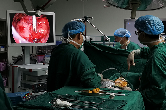
[Thoracoscopic Thoracic Surgery] 4K ultra-high definition thoracoscopy for major thoracic resection of lungs
Release time: 12 Dec 2023 Author:Shrek
Pulmonary bullae refer to air-containing sacs formed in the lung tissue due to various reasons causing the pressure in the alveolar cavity to increase, causing the alveolar walls to rupture and fuse with each other. There are two types of pulmonary bullae, congenital and acquired. Congenital disease is seen in children and adolescents. It is caused by congenital abnormal bronchial development, valve-like mucosal folds, and chondrodysplasia, causing valve action. Acquired disease is more common in adults and elderly patients, and is often accompanied by chronic bronchitis and emphysema. At present, the vast majority of bullous surgeries can be completed under the 4K ultra-high-definition thoracoscope camera system, and there is generally no recurrence after surgery. It is currently the best method to treat bullous pneumothorax.

Causes of bullae
It is generally caused by valve obstruction of small bronchi. It has the same formation mechanism as emphysema, but the degree is more severe.
The diameter of the alveoli of emphysema exceeds 1cm and occurs in the lung parenchyma. It is often accompanied by different lung diseases, such as bronchitis and bronchial asthma. It is also seen in patients without lung and bronchial diseases.
Due to inflammatory lesions and edema of the small bronchial mucosa, the lumen is partially blocked and produces a valve effect. After the air enters, it is difficult to discharge. The pressure in the alveoli increases, and the alveolar septum gradually ruptures, forming a huge air-containing sac cavity. This disease is clinically called this disease. .
Types of bullae
Type I: Narrow-necked bullae protrude from the lung surface and are connected to the lungs by a narrow band. This type of bullae has thin walls, is often formed by the pleura and connective tissue, and mostly occurs in the middle lobe.
Type II: Superficial bullae with broad bases, located on the surface of the lungs, between the visceral pleura and emphysematous lung tissue. Found in any part of the lungs.
Type III: Deep bullae with wide bases, similar to type II, but deeper and surrounded by emphysematous lung tissue. The bullae can extend to the hilus and can be found in any lung lobe.
Clinical manifestations of pulmonary bullae
The symptoms of patients with bullae are closely related to the number and size of bullae and whether they are accompanied by underlying lung diseases.
Small, small numbers of simple bullae may be asymptomatic and are sometimes discovered accidentally during chest X-ray or CT examination.
Large or multiple bullae may cause symptoms such as chest tightness and shortness of breath.
Sudden enlargement and rupture of pulmonary bullae can cause spontaneous pneumothorax, causing severe dyspnea, and chest pain similar to angina pectoris. Infection within the bulla may cause symptoms of pulmonary infection.
A small number of patients with bullae have symptoms such as hemoptysis and chest pain.
Thoracoscopic bullectomy is a minimally invasive surgery that can complete the entire bullae resection by making one or two small holes in the patient's chest wall on the affected side. A surgical camera is inserted into the chest cavity of the patient with lung bullae. The operator searches for and finds the lung bullae through thoracoscopic operating instruments. The operator uses a surgical stapler to remove the patient's lung bullae and remove them from the body to complete the entire operation. .
Compared with traditional "big surgery" surgery, the advantages of thoracoscopic bullectomy are that it takes a short operation time (only ten minutes), is minimally invasive (the wound is 2-3cm), and can basically achieve no bleeding. . It has less pain for patients, quick recovery after surgery, and low recurrence rate. It is now widely accepted by patients with pneumothorax and bullae.
The traditional "big knife"
The operation lasts more than 1 hour, involves large incisions, long recovery period, high cost, and great pain for the patient.
Minimally invasive thoracoscopic resection
The operation time is short, only more than 10 minutes, the wound is only 2-3cm, the patient suffers little pain, and the postoperative recovery is quick. Generally, the patient can be discharged from the hospital in 2-3 days.
(Single-port thoracoscopic bullectomy)
In summary, thoracoscopic bullectomy is now a very mature procedure. Nowadays, most bullous surgeries can be completed under video-assisted thoracoscopy, and 2/3 of patients' symptoms improve significantly after surgery. Those with better results were clearly demarcated and significantly enlarged bullae at the apex of the lung. Pulmonary function is basically not affected after subpleural bullectomy. For some patients with bilateral pulmonary bullae, minimally invasive surgery for bilateral pulmonary bullae can be performed using laparoscopy at the same time, with satisfactory surgical results.

- Recommended news
- 【General Surgery Laparoscopy】Cholecystectomy
- Surgery Steps of Hysteroscopy for Intrauterine Adhesion
- 【4K Basics】4K Ultra HD Endoscope Camera System
- 【General Surgery Laparoscopy】"Two-step stratified method" operation flow of left lateral hepatic lobectomy
- 【General Surgery Laparoscopy】Left Hepatectomy