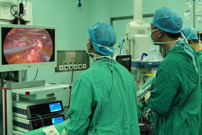
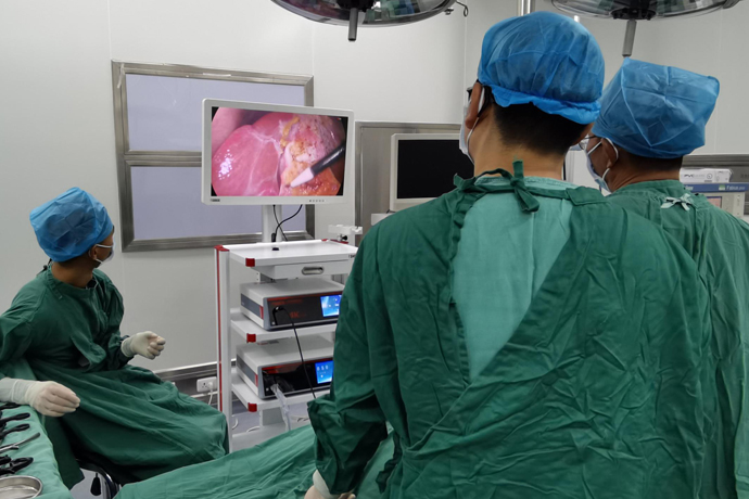
[General Surgery Laparoscopy] 4K Ultra HD Laparoscopic Left Hemihepatectomy
Release time: 03 Dec 2024 Author:Shrek
Left hemihepatectomy is more extensive than lobectomy. The left liver and right liver are divided into the diaphragmatic surface and the visceral surface with the gallbladder as the dividing point and the intra-abdominal great blood vessels as the dividing point. The scope of left hemihepatectomy is the left outer lobe and the left inner lobe. During this process, the gallbladder needs to be removed, the left hepatic duct, the left branch of the portal vein, and the left hepatic artery need to be cut off at the same time.

In addition, the left hepatic vein needs to be cut off at the second porta hepatis, which is the hepatic blood outflow tract, and only the middle hepatic vein should be retained, and the entire left liver should be resected. During hemihepatectomy, the risk is higher because the operation is performed around larger blood vessels, and there is a possibility of intraoperative massive bleeding. The operation needs to be performed in a better hepatobiliary center by a senior doctor with rich experience in liver resection. .
Left hemihepatectomy is more commonly used, especially for liver cancer and intrahepatic stones in the left lobe. The resection limit is about 0.5cm to the left of the median liver fissure, so as not to damage the middle hepatic vein that runs in the middle of the median liver fissure and joins the two middle liver lobes to return blood.
Preoperative diagnosis
1. Hepatolithiasis:
(1) Left liver regional type, the opening of the left hepatic duct is narrowed and obstructed, cholangitis.
(2) Left-sided Caroli disease.
2. Chronic cholecystitis
Operation time: April 28, 2020.
Surgical method: Laparoscopic left hemihepatectomy, Feiyanfu exploration surgery.
Surgical rules
Free liver.
The left hepatic pedicle was severed by extrathecal GIA.
On the basis of the hepatic ischemia interface, ultrasound is used to further confirm the projection of the middle hepatic vein.
The disconnection plane was determined based on the plane of hepatic ischemia, the course of the intrahepatic middle hepatic vein, and the plane of the canal of Arantius.
GIA severed the left hepatic vein.
Cholecystectomy, common bile duct exploration.
Trocar layout
Surgical Trocar layout and position:
Patient position: supine with legs apart, head high and feet low at 30 degrees.
Specific process:
1. Explore the abdominal cavity.
2. Free the liver.
3. Ligate the accessory left hepatic artery.
4. Place an blocking band on the first porta hepatis.
5. Dissect the left liver pedicle.
6. Intraoperative ultrasound determines the location of the middle hepatic vein.
7. Mark the liver resection plane according to the ischemic interface.
8.Pringle method blocks the first portal of liver.
9. Cut the liver along the marked line.
10. Cut off the first IVB branch of the middle hepatic vein.
11. Cut off the second IVB branch of the middle hepatic vein.
12. Cut off the first branch of the middle hepatic vein IVA.
13.GIA separates the left hepatic vein.
14. Release the block.
15. Hemostasis of liver section.
16. Remove the gallbladder.
17. Biliary tract exploration.
18. Place T-tube on the side wall.
19. Remove the specimen.

- Recommended news
- 【General Surgery Laparoscopy】Cholecystectomy
- Surgery Steps of Hysteroscopy for Intrauterine Adhesion
- 【4K Basics】4K Ultra HD Endoscope Camera System
- 【General Surgery Laparoscopy】"Two-step stratified method" operation flow of left lateral hepatic lobectomy
- 【General Surgery Laparoscopy】Left Hepatectomy