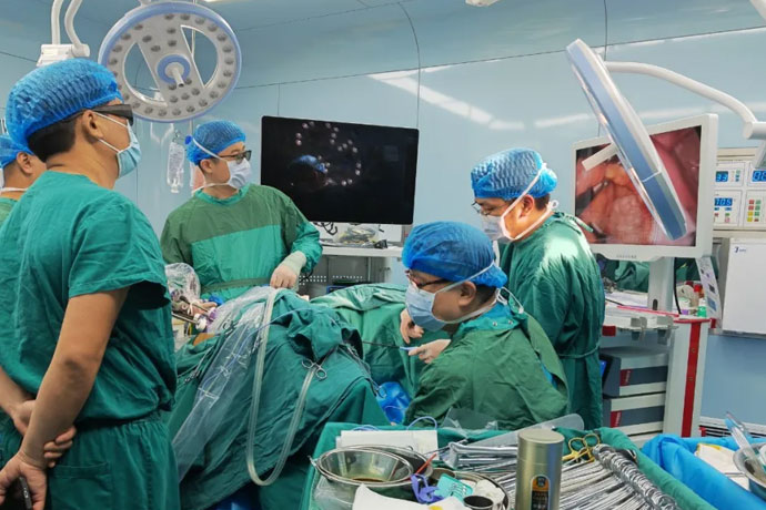
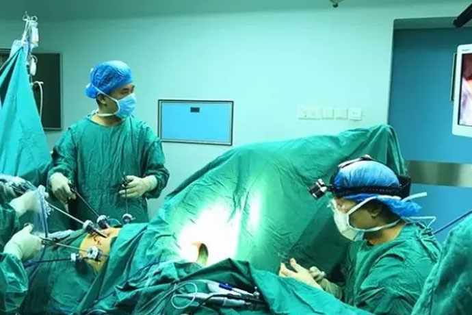
[General Surgery Laparoscopy] 4K Ultra HD Laparoscopic Retrorectal Suspension Patch Fixation
Release time: 24 Dec 2024 Author:Shrek
Rectal prolapse (RP) refers to the downward displacement of the anal canal, rectum, and even the lower end of the sigmoid colon, protruding outside the anus, and is accompanied by pelvic floor dysfunction. Although rectal prolapse is a benign disease, the discomfort, mucus exudation and bleeding caused by the prolapse, as well as the accompanying fecal incontinence or constipation, will seriously affect the patient's quality of life. Women over 50 are 6 times more likely to develop the disease than men. Although rectal prolapse is generally thought to be related to childbirth, nearly one-third of female patients have no history of childbirth. About 50% to 75% of patients with rectal prolapse have anal incontinence, and 25% to 50% of patients have constipation. Half of the patients have pudendal neuropathy, which may be due to denervation and atrophy of the external anal sphincter.

Domestic classification of external rectal prolapse has been detailed, including:
Grade I prolapse is about 3 cm in length during defecation and can retract on its own after defecation.
Grade II prolapse is full-thickness prolapse of the rectum during defecation, with a length of 4 to 8 cm and must be reduced by manual pressure.
Grade III prolapse refers to the prolapse of the anal canal, rectum and part of the sigmoid colon during defecation, with a length of more than 8 cm and is difficult to reduce.
Conservative treatment methods such as traditional Chinese medicine, biofeedback, and injection therapy can be used for grade I rectal prolapse; transanal surgery can be used for grade II rectal prolapse; transabdominal surgery is preferred for grade III rectal prolapse, and the purpose of surgical treatment is to cure anatomic abnormalities. , improve the accompanying symptoms of fecal incontinence, constipation and pain.
Rectal prolapse is a disease in which the rectum is fully intussuscepted and prolapses outside the anus. It often occurs together with the following anatomic abnormalities, such as levator ani muscle laxity, excessively deep Douglas fossa, long sigmoid colon, hole-like anal sphincter, and sacrorectopexy. lost or weakened.
The advantages of laparoscopic retrorectal suspension patch fixation are:
1. Follow the principle of protecting the pelvic autonomic nervous system (PANP) during laparoscopic radical resection of rectal cancer, and preserve the pelvic autonomic nervous function to the greatest extent under direct vision, especially focusing on protecting the neurovascular bundles (NVB) on both sides of the anterior wall. The patient's urinary function and sexual function are protected.
2. Laparoscopic suturing of the patch on the sacral promontory, suturing of the patch to the "peritoneal wings" on both sides, and encircling suturing to eliminate the recto-vesical depression and elevate and reconstruct the pelvic floor ensure a low postoperative recurrence rate.
3. This surgical operation is completed under laparoscopy, with few and small abdominal wall incisions, mild postoperative incisional pain, good cosmetic effect, few complications, and high patient acceptance.
Establish operating platform
After successful general anesthesia, the lithotomy position was adopted, and the surgical field was routinely disinfected and draped.
1. Make a circumumbilical incision in the skin fold above the umbilicus, about 1.2cm long, and enter the abdomen layer by layer. A 12mm Trocar was inserted through the incision, pneumoperitoneum was established and the air pressure was maintained at 13-15mmHg. A 30° laparoscopic lens is inserted through this Trocar.
2. Under the observation of the laparoscopic monitor, avoid the inferior epigastric artery and insert a 5mm Trocar on the line connecting the lateral edge of the right rectus abdominis and the horizontal line of the umbilicus as the auxiliary operating port of the main surgeon.
3. Insert a 12mm Trocar into the McBurney's point on the right side as the main operating hole for the main knife.
4. Insert a 5mm Trocar at the intersection of the lateral edge of the left rectus abdominis and the horizontal line of the umbilicus as the assistant's main operating hole.
5. Insert 5mm Trocar into the anti-McFarland point on the left side as the assistant's auxiliary operating hole.
Explore
No abnormalities were found in the liver, gallbladder, stomach, duodenum, jejunum, cecum, ascending colon, transverse colon, and descending colon; the inherent adhesion zone between the lateral edge of the first curve of the sigmoid colon and the left abdominal wall was missing, and no intersigmoid saphenous was found. nest. The intraoperative diagnosis was consistent with the preoperative diagnosis, and it was decided to perform "4k laparoscopic retrorectal sling + patch fixation".
Surgical steps
1. Medial dissection of the left colon:
(1) Incise the midline side of the sigmoid mesocolon: Use intestinal forceps to grasp the rectum and pull it ventrally, tighten the sigmoid mesocolon, use the sacral promontory as the entry point, and use the "yellow-white junction line" as a guide, from the caudal side to the cephalad side. After incising the mesocolon to the root of the small intestinal mesentery and turning left, a loose gap can be seen, which is the fused fascial space (Toldt space) between the left mesocolon and the prerenal fascia.
(2) Expand the Toldt space: the assistant's intestinal forceps continue to pull the upper rectum ventrally, and the right intestinal forceps grasps the inferior mesenteric artery pedicle toward the head and maintains tension ventrally. The surgeon carefully expands the Toldt space: within this space, expand the surgical operation to the left side. plane, reaching Toldt's line where the sigmoid mesocolon disappears. Attention was paid to maintaining the integrity of the left mesocolon and prerenal fascia, and retaining a layer of transparent prerenal fascia in front of the main iliac vessels. Through this fascia, the left ureter and reproductive vessels can be seen posterolaterally at the root of the sigmoid mesocolon. (No damage was caused to the inferior mesenteric plexus, left ureter, and left reproductive blood vessels). The separation range was from the center to the left to the left paracolic groove lateral to the reproductive blood vessels, and from the caudal side to the head side to the root of the inferior mesenteric artery.
2. Posterolateral dissection of left colon:
Traction the sigmoid mesocolon to the right, starting from the inherent adhesion zone between the lateral edge of the first curve of the sigmoid colon and the left abdominal wall, and incise the peritoneal reflection of the left paracolic groove cephalad along the yellow-white junction line (Toldt line). The sigmoid colon was turned to the right, and the Toldt space between its mesentery and prerenal fascia was mobilized to the right. Care was taken to protect the left ureter and left genital blood vessels behind the prerenal fascia from damage. The lateral sigmoid colon was completely connected to the midline lateral plane, and extended upward to the level of the lower sigmoid colon. Care was taken to protect the integrity of the prerenal fascia, sigmoid mesocolon, and original descending mesocolon.
3. Perirectal mobilization:
Starting from the level of the sacral promontory, in the loose connective tissue space behind the upper mesorectum, the surgical plane is sharply extended posteriorly and laterally to the retrorectal space and to the supralevator ani space.
(1) Posteriorly: starting from the level of the sacral promontory, close to the mesocolorectum, expand the surgical plane caudally in the retrorectal space between the mesocolorectum and the presacral fascia, cut off the rectosacral fascia, and enter the levator ani Supramuscular space, close to levator ani muscle.
(2) Laterally: Expand the retrorectal space to both sides, and use the posterior space as a guide to free the perirectal space to both sides until the level of the suprarectal space.
The inferior mesenteric artery and superior rectal artery were not cut, the rectal ligaments were not severed, and the peritoneal reflection in front of the rectum was not opened. During the whole process, attention was paid to protecting the integrity of the pelvic autonomic nerves.
4. Retrorectal sling fixation:
Choose a piece of hernia mesh (material: polypropylene), trim it to a suitable shape, and place it behind the rectum in the abdominal cavity. Make sure that the mesh is fully expanded and laid flat in the gap behind the rectum.
After the assistant lifts the rectum and corrects the prolapse, the upper section of the patch is fixed to the anterior fascia of the sacrococcygeal promontory with absorbable sutures (note: it must be fixed firmly! Looseness is not allowed), embed the patch, and carefully Stop bleeding (to prevent postoperative pelvic floor infection!); Use absorbable 3-0 Vicryl sutures on the left side of the rectum to wrap and bury the left peritoneum, patch, and left rectal fascia to prevent the patch from contacting the rectal surface. Complete peritonealization (purpose: to avoid adhesion between the small intestine and the patch).
Finally, the left lateral seromuscular layer of the sigmoid colon was sutured to the left lateral peritoneum to suspend and fix the sigmoid colon to the lateral abdominal wall.
5. Postoperative examination
The surgical field was cleaned, the abdominal cavity was repeatedly flushed with normal saline, and the suction was performed. Check that there is no active bleeding in the abdominal cavity. Reposition the intestinal tube, count the instruments and gauze points, and observe that there is no bleeding in each puncture hole. A 26-gauge chest drainage tube was inserted into the operating hole in the right lower abdomen to the pelvic floor and fixed. The laparoscope and each puncture cannula were withdrawn, each operating hole was sutured layer by layer, and covered with a small dressing.

- Recommended news
- 【General Surgery Laparoscopy】Cholecystectomy
- Surgery Steps of Hysteroscopy for Intrauterine Adhesion
- 【4K Basics】4K Ultra HD Endoscope Camera System
- 【General Surgery Laparoscopy】"Two-step stratified method" operation flow of left lateral hepatic lobectomy
- 【General Surgery Laparoscopy】Left Hepatectomy