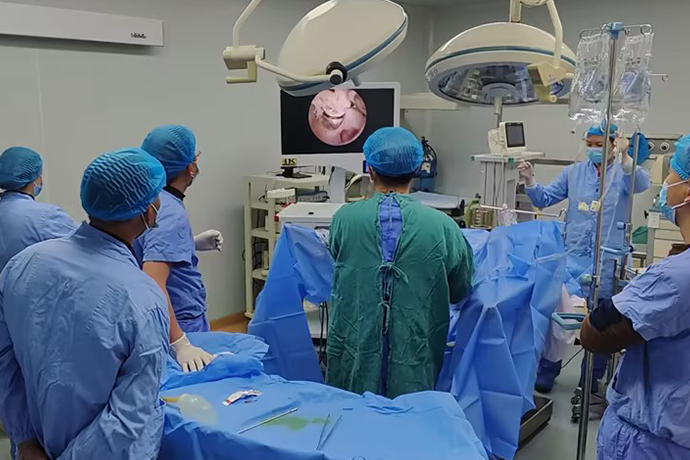
[Gynecological laparoscopy] Causes and treatments of urinary system injuries
Release time: 11 Jan 2025 Author:Shrek
The anatomical relationship between the female reproductive system and the urinary system is closely adjacent. During gynecological surgery, due to the invasion of malignant tumors, severe adhesions of pelvic organs, or large tumors in the pelvic and abdominal cavity, variations in the anatomical position of the urinary system and unclear tissue levels can occur. Can easily cause damage to the urinary system. Although there is currently no systematic review comparing the incidence of urinary tract injuries between open and laparoscopic surgery.

In clinical practice, more and more benign and malignant gynecological surgeries can be completed under laparoscopy. However, with the expansion of gynecological laparoscopic surgery applications and the increase in surgical difficulty, the incidence of intraoperative and postoperative complications has also shown an upward trend. Severe irreversible damage will have a serious impact on patients' postoperative quality of life.
Causes of Urinary System Damage
Urinary tract injury is one of the most common complications of laparoscopic surgery.
Since laparoscopic surgery often uses electrocoagulation and electrocution to treat blood vessels and tissues, the local temperature can be as high as 300°C. Especially when a single-stage electric hook is used to perform electrocoagulation and resection near the ureter, the heat conduction effect can spread as far as the surrounding area. 2cm range, causing ischemic necrosis of the ureter. This kind of thermal injury is often not easy to detect during the operation, but the corresponding symptoms and signs appear for a period of time after the operation. Once an abnormal increase in drainage volume is found, the possibility of urinary system fistula should be considered. It is usually considered in two steps. First, it is necessary to determine whether there is a urinary system fistula, and secondly, it is necessary to determine the location of the fistula.After the operation, the creatinine and urea nitrogen indicators of the pelvic drainage fluid can be checked to determine whether there is a urinary system fistula; the bladder methylene blue test can be used to determine whether it is a vesicovaginal fistula or a ureterovaginal fistula; further cystoscopy and CT urinary tract imaging (CTU) are performed. The side of the injury and the location of the fistula can be clarified. It is necessary to pay attention to whether there are compound fistulas, that is, there are ureteral fistulas and bladder fistulas at the same time, bilateral ureteral fistulas, and more than two bladder fistulas.
Intraoperative and postoperative management of ureteral injury
When the tubular cord is found to be cut or injured during the operation, or there is a lot of exudate in the surgical field, the source of the exudation should be carefully searched to identify the ureteral injury. If it is confirmed that the ureter has been accidentally tied or clamped, it should be removed immediately. If the clamping or suturing time is short and the damage is not obvious, the ureter can be left for a period of time to observe the ureteral peristalsis. The ureteral peristalsis is good and does not need to be treated. If the injury is obvious and the peristalsis function is lost, it should be treated as appropriate. In mild cases, a double "J" ureteral catheter can be placed. In severe cases, end-to-end ureteral anastomosis or ureterovesical anastomosis can be considered after cutting off.
If the ureter is found to be severed or seriously injured during the operation, end-to-end ureteral anastomosis can be performed if the position is higher. Drainage is placed next to the anastomosis. At the same time, a double "J" ureteral catheter is placed in the ureter, and the upper end to the lower end of the renal pelvis is placed in the bladder. The catheter was removed via cystoscopy 2 weeks after surgery. If the location of the injury is low, ureterovesical anastomosis can be performed, and a double "J" ureteral catheter can also be placed, and the catheter can be removed through a cystoscope 2 weeks later.
Different treatment methods are selected according to the patient's general condition, injury site, defect range, local blood supply, infection, etc. Patients with no obvious obstruction to the IVU, smooth retrograde intubation, no chills, fever, and little urine leakage can be treated. Built-in double "J" tube drainage and anti-infective conservative treatment for 10-14 days. Once ureteral dilatation occurs, ureterovesical reimplantation or other treatments should be performed promptly to save renal function. For those with serious injuries, the injured part should be resected decisively, an end-to-end anastomosis of the ureter should be performed or it should be transplanted into the bladder and double ""J tubes should be placed for 4-6 weeks. For early ureteral fistulas with small postoperative urinary fistulas, ureteral catheters can be placed for continuous drainage for 10-14 days, and healing is possible. When it is difficult to place a large fistula or double "J" and conservative treatment is ineffective, a ureteral stent or nephrostomy is performed, and a second-stage repair is performed 3 to 6 months after the operation.
Characteristics, prevention and treatment of bladder injuries
1. Features
The incidence rate of bladder injury is about 0.34%, and the injury locations are mainly the top, bottom and trigone of the bladder. Causes of injury include: 1) poor technique and unclear anatomical levels; 2) thermal damage to instruments [6; 3) various factors leading to local adhesion, such as cervical tumor infiltration, history of cesarean section, endometriosis, reproductive Systemic tuberculosis, inflammatory reactions, etc. not only increase the difficulty of surgery, but also increase the probability of bladder damage during the separation of the bladder, cervix, and vaginal space1. Hong et al[? retrospectively analyzed 118 cervical cancer patients who underwent laparoscopic radical hysterectomy. Bladder injury occurred in 10 cases (8.47%), 5 of which had a history of cesarean section, resulting in damage to the bladder base and bladder trigone.
2. Prevention
2.1 Anatomy
The bladder is composed of mucosal layer, muscular layer and adventitia, and the outer membrane surface is covered by peritoneum. The peritoneum is tightly connected to the adventitia at the midline of the bladder tip and upper body, but is loosely connected to the adventitia on the side of the bladder body, making the peritoneum easy to peel off. This is the anatomical basis for extraperitoneal dissection to reach the lower uterine segment. There is a thin layer of loose connective tissue gap between the bladder and cervix, located between the trigone of the bladder and the cervix. Its upper boundary is the vesicouterine reflex peritoneum, its lower boundary is the superior vaginal septum, and both sides are the medial leaves of the vesicocervicovaginal ligament. The gap can be seen as a downward continuation of the bladder-cervical gap. There are few blood vessels in it and it is easy to separate.
2.2 Precautions
In patients with no obvious adhesions in the lower uterine segment, open the bladder and uterus and fold the peritoneum, lift the uterine cup with force to expose the bladder, cervix and vaginal space, gradually push the bladder down along the gap, and separate and expand downward and on both sides to achieve enough space for surgery. level. If the level of adhesion is unclear and the anatomical level of the connective tissue gap between the bladder and cervix has not been clarified, an ultrasonic scalpel can be used to sharply separate the cervix as close as possible, combined with a blunt suction device to gently push down to feel the push-down tension, resistance and bleeding. If the tension and resistance are high and bleeding is easy, it often means that the anatomical level is not good. At this time, you should not force the separation to avoid bleeding. You should separate sharply and find the correct boundaries, and then perform blunt separation v to avoid forced blunt separation that may cause bladder muscle damage. Layer blood vessel damage, bleeding or even bladder rupture; such as tension Or the resistance is small and the bleeding is very little, it often means that the anatomical level is correct, and the white and smooth bladder and cervical fascia can be seen. At this time, the blunt push down of the suction device will be more effective, and it will stop when the resistance increases, and then sharp If the separation is repeated, the bladder can be pushed down with less bleeding. For some patients with adhesions between the bladder and the lower uterus, you may consider opening the lateral bladder fossa first, bluntly and sharply separating the adhesions from the side of the bladder via an extraperitoneal approach, and then pushing down the bladder. However, attention should be paid to the anatomy of the lateral bladder fossa and do not damage the lateral wall of the bladder.Although there is no exact mechanism for fistula formation, experimental studies in animals by Cogan et al. have shown that the formation of vesicovaginal fistula after laparoscopic hysterectomy is related to unipolar thermal damage, so it is best to use ultrasonic scalpel when resecting the vaginal wall. Replaces monopole, reducing the possibility of thermal damage.
3. Processing
After the bladder is separated and moved, a timely examination should be carried out. If bladder damage is found, it should be repaired immediately. If there is a high degree of suspicion but no obvious damage can be seen, intraoperative injection of methylene blue solution or cystoscopy can be used to help diagnose R1. Only muscle layer injuries can be sutured with 4-O absorbable synthetic sutures intermittently. If the break has been penetrated, two layers of suturing should be performed. After full-thickness suturing with 4-0 absorbable suture, one layer of sutures should be inverted and sutured; the break is located in the trigone of the bladder. When suturing near the ureteral area, be careful not to injure the ureteral opening; adequate bladder drainage for 2 weeks after surgery and prevention of infection can usually lead to primary healing.Most electrical injuries to the bladder are difficult to detect during surgery, usually manifesting as a large amount of vaginal discharge about one week after the operation or a small amount of discharge at first but gradually increasing and causing infection. At this time, the patient should actively inject methylene blue solution into the bladder and perform retrograde treatment. Cystoureterography, cystoscopy, B-ultrasound, creatinine, urea nitrogen, electrolyte testing and comparison of vaginal exudate and urine were performed to determine whether there was a urinary fistula and its location. As soon as it is discovered, an indwelling urinary catheter should be placed immediately for adequate bladder drainage to control urinary tract infection, and the patient should try to adopt a position that can reduce urinary fistula exudate. Most bladder injuries and vesicovaginal fistulas can be healed in the first stage without leaving sequelae. For vesicovaginal fistulas that are unsuccessfully conservative, secondary repair is usually performed 3 to 6 months after surgery.

- Recommended news
- 【General Surgery Laparoscopy】Cholecystectomy
- Surgery Steps of Hysteroscopy for Intrauterine Adhesion
- 【4K Basics】4K Ultra HD Endoscope Camera System
- 【General Surgery Laparoscopy】"Two-step stratified method" operation flow of left lateral hepatic lobectomy
- 【General Surgery Laparoscopy】Left Hepatectomy