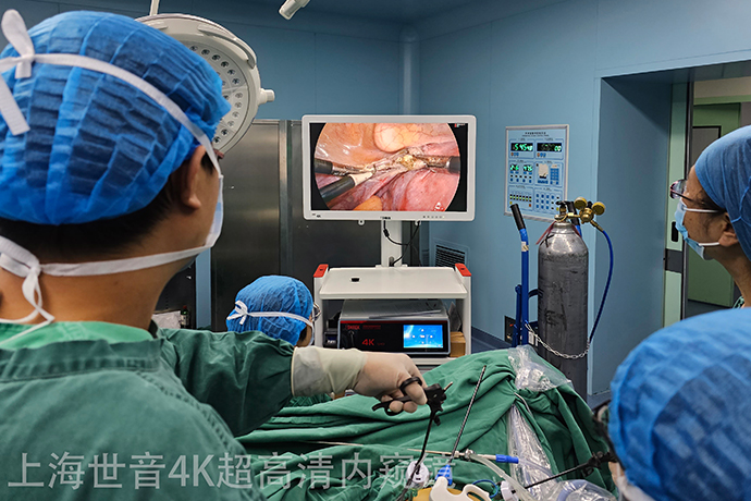
[Gynecological Laparoscopy] 4K UHD Laparoscopic Myomectomy
Release time: 20 Jun 2023 Author:Shrek
Now the concept of health examination is becoming more and more popular. The "uterine fibroids" that often appear in the medical examination reports make female friends extremely worried. When they hear the word "tumor" in their names, they start to worry, especially when they see the "uterine fibroids" in the report. The described fibroids are huge in size, and I am even more confused. I am afraid of surgery, and I don’t know what to do?

What are uterine fibroids?
It is the most common benign tumor of the female reproductive system. It is composed of smooth muscle and connective tissue. It is common in women aged 30-50. About 20% of women over the age of 30 have uterine fibroids. Don't look at the word "tumor" in the name, it is kind in nature, and the rate of malignant transformation (also called sarcoma-like transformation) is only 0.4%-0.8%.
Why do uterine fibroids develop?
The exact cause is unknown, and it may be related to female hormones. Therefore, it is rare before puberty, and it is prone to growth and denaturation during pregnancy. It stops growing or shrinks after menopause, and it may also be related to genetics.
The uterus is the "house" where the fetus is born.
(1) Submucosal fibroids: Fibroids grow into the "room", which is a foreign body in the room, which will affect the internal structure of the room, and is prone to symptoms of increased menstrual flow and prolonged menstruation, leading to anemia.
(2) Intramural fibroids: grow in the "wall". If the wall is thick enough, the fibroids are small and will not cause symptoms. When these fibroids grow in size and protrude into the room, they may appear similar to submucosal muscle tumors. Symptoms of tumor, if it protrudes to the outside of the room, it will not have much impact.
(3) Subserosal fibroids: They grow outside the "room" and are generally asymptomatic. If they grow up to oppress adjacent organs such as the bladder and rectum, corresponding symptoms such as frequent urination, dysuria, and constipation may appear. If the fibroid grows to the left/right side of the room and protrudes between the two leaves of the broad ligament, it is a "broad ligament fibroid".
(4) Cervical fibroids: As the name suggests, they grow on the cervix, and the growth site is relatively low, close to the surrounding blood vessels, ureter, etc., with rich blood supply, which may lead to unclear surrounding anatomy and compression symptoms.
What are the symptoms of uterine fibroids?
Answer: Most uterine fibroids grow "quietly" without symptoms, and are found during physical examination. Therefore, we remind all female friends to have regular physical examinations. Its symptoms are not related to the location, size, degeneration and number of fibroids.
(1) Menstrual changes: the most common, submucosal fibroids and larger intramural fibroids may cause increased menstrual flow, prolonged menstrual periods, and even lead to anemia.
(2) Mass in the lower abdomen: When the fibroids enlarge and the uterus exceeds the size of 3 months of gestation, the mass will become palpable.
(3) Symptoms of compression: Fibroids grow forward, backward, and left and right, which may oppress surrounding organs and blood vessels, resulting in corresponding compression symptoms, such as frequent urination, dysuria, urinary retention, constipation, hydroureter, lower extremity edema, etc. .
(4) Others such as increased leucorrhea, lower abdominal distension, back pain, infertility or miscarriage.
What is the best way to do surgery?
At present, surgery is the main treatment method. Through 4K ultra-high-definition laparoscopic surgery for uterine fibroids, the surgical screen has 3840*2160 ultra-high-definition pixels, and doctors can find the location of fibroids more accurately. The abdominal incision is small, the postoperative pain is less, the recovery is faster, and the hospitalization time of the patient is shortened. Under the magnified field of view of the 4K ultra-high-definition laparoscope, the surgical operation is more precise, and the incidence of complications such as abdominal and pelvic adhesions and intestinal adhesions after the operation is reduced. The mainstream surgical method for the treatment of uterine fibroids.
The same is uterine fibroids, what you look like may be very different from what others look like. According to its relationship with the muscle layer, uterine fibroids can be roughly divided into three categories: submucosal fibroids, intramural fibroids, and subserosal fibroids.
Submucosal fibroids
Submucosal fibroids grow under the endometrium, which is a type of mucosa, so the surface of this fibroid will be covered with a layer of mucous membrane.
This will virtually increase the bleeding area during menstruation, which will easily lead to heavy menstrual flow, long menstrual periods and even anemia.
In addition, if the fibroids are relatively large, the uterine contraction will compress the fibroids during menstruation, resulting in dysmenorrhea.
There is also a rather special fibroid with a particularly long pedicle, some of which can even grow to the outside of the vagina, and it will be exposed when the patient squats down, which is quite scary.
Subserosal myoma
Subserosal fibroids most of the time do not have any symptoms. However, if the fibroids are relatively large and oppress other surrounding organs, it will cause corresponding symptoms.
For example, compression to the bladder may cause frequent urination and urgency; compression to the rectum may cause tenesmus or constipation; in rare cases, compression to the ureter may cause ureteral obstruction, resulting in hydrops in the kidneys.
Intramural fibroids
If the intramural fibroids are particularly large and numerous, it will cause uterine deformation, and the endometrium may also bulge, resulting in heavy menstrual flow or prolonged menstruation.
Laparoscopic surgery steps
The general steps of laparoscopic surgery include the following:
1. Use a Veress needle to infuse the stomach with carbon dioxide gas to inflate the abdominal cavity.
2. Make 3-4 incisions of 5-10mm on the belly.
3. A uterine lifter that can move the uterus should be placed in the vagina, the main purpose is to assist in the adjustment of the position of the uterus during surgery (even some patients who have no sex life need to lift the uterus, this step is very important).
4. Inject diluted pituitrin on the surface of the uterus to reduce intraoperative bleeding.
5. Cut the surface of the uterus to expose the fibroids, and use laparoscopic instruments to completely separate the fibroids.
6. Suture the uterine incision back with absorbable suture.
7. Use a fibroid rotary cutter to crush the fibroids and take them out from the wound on the abdominal wall. The diameter of the rotary cutter is usually 10-15mm. At this time, the abdominal wall incision needs to be extended to 10-15mm.
8. To prevent adhesions, place an anti-adhesion film on the surface of the uterus.
9. Take out the instrument and suture the wound.
Uterine fibroids are the most common tumors of the female reproductive system, mostly in women aged 35 to 50. Uterine fibroids are generally a few centimeters in size. Such huge uterine fibroids are very rare. The larger the tumor, the greater the risk of surgical removal. Once malignant transformation or hemorrhage occurs, it will pose a danger to the patient's life. Here, female friends should have a regular gynecological examination every year. If gynecological diseases such as uterine fibroids are found, they should be treated as soon as possible to ensure life safety and health.
Postoperative precautions
1. Laparoscopic surgery is a minimally invasive surgery, so you can turn over on the bed 6 hours after the operation, and get out of bed 24 hours after the operation, so as to prevent intestinal adhesions.
2. You need to rest for 1 month after the operation and try to avoid lifting heavy objects or doing strenuous sports.
3. Sexual life and bathing are prohibited within 1 month after surgery.
4. Color Doppler ultrasound should be checked one month after operation to see how the uterus is recovering.
5. Take contraceptive measures within two years after the operation, and try not to get pregnant, because after all, there will be scars on the uterus after the operation. If you get pregnant early and have an abortion, it may lead to uterine perforation, and there is also a risk of uterine rupture in the third trimester, so It is safer to conceive again two years after the operation.

- Recommended news
- 【General Surgery Laparoscopy】Cholecystectomy
- Surgery Steps of Hysteroscopy for Intrauterine Adhesion
- 【4K Basics】4K Ultra HD Endoscope Camera System
- 【General Surgery Laparoscopy】"Two-step stratified method" operation flow of left lateral hepatic lobectomy
- 【General Surgery Laparoscopy】Left Hepatectomy