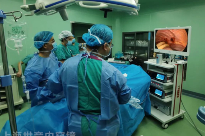
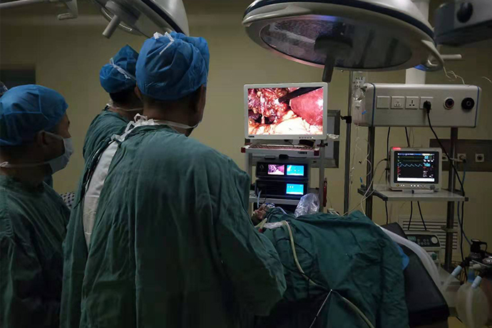
[Laparoscopy in Hepatobiliary Surgery] Incision and drainage of liver abscess
Release time: 13 Jun 2023 Author:Shrek
Liver abscess is a disease in which bacteria or other pathogens invade the liver parenchyma or bile duct, triggering a local inflammatory response, which in turn leads to the formation of an abscess inside the liver. It mainly includes bacterial liver abscess and amebic liver abscess, which is a serious secondary infection of the liver, among which bacterial liver abscess is relatively more common in clinical practice.
Liver abscess is a purulent lesion of the liver with a mortality rate of up to 10%. Patients with liver abscess often present with irregular intermittent high fever, accompanied by a history of diarrhea or diabetes.
Physical examination revealed enlarged liver, localized edema and tenderness in the intercostal space, and occasional jaundice. If an abscess is perforated, empyema or peritonitis will result.

Common types of liver abscess
1. Bacterial liver abscess accounts for 80%, showing multiple small abscesses, or multiple small abscesses fused into a large abscess.
2. About 10% of amebic liver abscesses are solitary.
3. Fungal liver abscess 10%.
The ducts in the liver include the biliary tract, portal vein, hepatic artery and vein, and lymphatic vessels. When puncturing a liver abscess, care should be taken to avoid important ducts.
Causes of liver abscess
1. Biliary tract disease: The existence of diseases that can cause obstruction in the biliary tract, such as bile duct stones, biliary roundworms, and biliary tract tumors, can lead to poor bile drainage and secondary bacterial infection, leading to the occurrence of liver abscess.
2. Intra-abdominal infection: If there is an infection focus in the abdominal cavity, the microorganisms in the focus can enter the liver along with the portal vein blood flow, causing local inflammation of the liver.
3. Infection in organs adjacent to the liver: when anatomically similar to the liver, an organ is infected or inflamed, the infection focus may directly spread and invade the liver due to the close distance, leading to infection.
laparoscopic approach
Laparoscopic abscess incision and drainage: the purpose is to incise the liver abscess and drain the pus in a minimally invasive surgical method, which helps to quickly relieve symptoms and promote disease recovery.
Take the head high and feet low, pneumoperitoneum pressure 13~15mmHg, place a 10mmTrocar on the navel (point A) and place a laparoscope, place a 5mmTrocar on the anterior axillary line of the right costal margin (point E), and use a pneumoperitoneum needle on the intercostal or abdominal wall under the laparoscope Puncture the lowest part of the corresponding abscess. Take out the pus to judge the depth, maturity and nature of the abscess. If the pus is accompanied by bile, an exploratory laparotomy is required to prevent the abscess from rupturing into the biliary tract. After confirming that the abscess has matured, a 5mm Trocar (point F) was disposed at this point, and care should be taken to avoid the incision here from entering the thoracic cavity and causing pneumothorax (all were positioned by B-ultrasound before operation). Subphrenic adhesions can be peeled off with an electrocoagulation hook to reveal the abscess. First, a small hole is opened with the electrocoagulation hook at the highest point or thinnest part of the abscess to allow the pus to flow out, and the pus is sucked up, and then the suction tube is inserted into the abscess cavity to continue suctioning. , Gently turn the suction device to separate the abscess cavity until the pus is completely sucked, carefully clean the pus moss, and rinse the abscess cavity with hydrogen peroxide or metronidazole and normal saline. At the end of the operation, a silicone drainage tube was placed in the abscess cavity and around the liver, and fixed on the abdominal wall.
Postoperative routine intravenous infusion of broad-spectrum antibiotics for 3 to 5 days, and active symptomatic and supportive treatment, and then selected sensitive antibiotics for 10 to 14 days according to the results of pus culture and drug sensitivity test, until the body temperature was normal for 3 days, and the symptoms disappeared and then oral antibiotics were used. The abscess cavity can be flushed through the abscess cavity drainage tube, and the drainage fluid can be removed if the drainage fluid is less than 20ml per day. Generally, the drainage tube in the abscess cavity is removed 2~4 days after the operation, and the abdominal drainage tube is removed 1~2 days later, so as to observe the abscess Whether there are complications such as bile leakage or bleeding after lumen extubation, and residual pus that may flow out.
Precautions
①The adhesion between the liver and the abdominal wall or diaphragm is the liver abscess. The adhesion is mostly loose, and the electrocoagulation hook is easy to separate. The incision range of the electrocoagulation hook should reach the adhesion boundary.
②Gauze strips should be placed on the upper and lower poles of the abscess before incision of the liver abscess to prevent the spread of contamination.
③ Generally, the operation is performed 1 week after the onset, when the abscess has matured and liquefied.
④ When separating the septum of the abscess cavity, if the tubular frame structure of the liver is seen, there is no need to dissect it. During the dissection, the abscess wall should not go beyond the wall of the abscess cavity to normal tissue. It is found that pulsating bleeding with titanium clips can effectively prevent complications such as bleeding and bile leakage.
⑤ It is best to install a double-chamber drainage tube for the abscess cavity, which is convenient for postoperative flushing, and avoids the thorax and hepatic hilum.

- Recommended news
- 【General Surgery Laparoscopy】Cholecystectomy
- Surgery Steps of Hysteroscopy for Intrauterine Adhesion
- 【4K Basics】4K Ultra HD Endoscope Camera System
- 【General Surgery Laparoscopy】"Two-step stratified method" operation flow of left lateral hepatic lobectomy
- 【General Surgery Laparoscopy】Left Hepatectomy