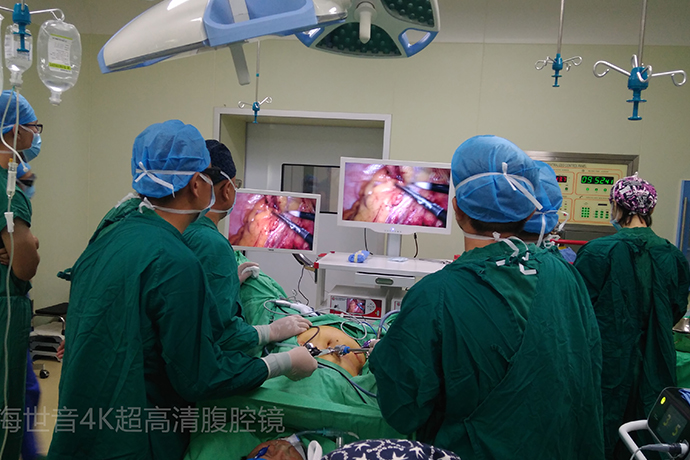
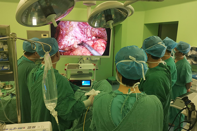
[General Surgery Laparoscopy] 4K ultra-high definition laparoscopic sigmoidectomy
Release time: 07 May 2024 Author:Shrek
What is sigmoid colon cancer?
Sigmoid colon cancer is a type of colon malignant tumor that occurs in the section of colon between the descending colon and the rectum. Most of the early symptoms are mild or not obvious and are easily ignored. Symptoms include abdominal pain, indigestion, and bloating. In the middle and late stages, the clinical manifestations include persistent abdominal discomfort, dull pain, bloating, constipation, and blood in the stool. The symptoms cannot be relieved after general treatment. Symptoms of intestinal obstruction may occur.

Colon cancer is a common malignant tumor of the digestive tract. According to the latest data on malignant tumor diseases in China released by the National Cancer Center, colorectal cancer has ranked second in the incidence of malignant tumors, seriously threatening patients' lives and health. If not treated in time, it may Leading to a serious decline in the patient's quality of life or even death. The treatment of colon cancer is still mainly surgical. Surgery is currently the only treatment method that can cure colon cancer. In recent years, with the advancement of medical technology and the combination of surgery and postoperative chemotherapy and other adjuvant treatments, the five-year survival rate and cure rate of colon cancer patients have steadily increased. Early detection and early treatment of colon cancer are key factors affecting the prognosis of patients. However, the early symptoms of this disease are not obvious, so if you find changes in bowel habits, abdominal masses, and some unexplained anemia, weight loss and other symptoms in daily life, , it is recommended to seek medical treatment in a regular medical institution as soon as possible. In addition, current relevant guidelines recommend that people over the age of 50 undergo routine colonoscopy every year to achieve early detection and early treatment.
key step
1. Cannula position: Use the Hassion method to place a 10mm trocar in the umbilicus, a 12mm trocar in the right iliac fossa, a 5mm trocar in the right upper abdomen, and a 5m trocar in the left iliac fossa (optional).
2. Adjust the operating table so that the patient tilts to the right and assumes a mild head-down position.
3. Laparoscopic exploration, place the small intestine and omentum in the right upper abdomen.
4. Confirm and separate the inferior mesenteric vascular pedicle to protect the ureter and presacral autonomic nerve.
5. Release the descending mesocolon in the retroperitoneum.
6. Dissect the lateral attachment of the sigmoid colon and descending colon (peritoneum) to the splenic flexure.
7. Free the rectosigmoid-intestinal junction and select the transverse section.
8. Transect the upper rectum and its mesentery.
9. Remove the specimen through the left lower abdominal incision and remove the sigmoid colon.
10. Reconstruct pneumoperitoneum and parallel (colorectal) anastomosis.
11. Close the incision.
Patient position
The patient lies supine on the bean-shaped inflatable bag on the operating table. After successful induction of general anesthesia, a gastric tube and a urinary tube are placed, and the legs are fixed on Dan Allen or Yellowfin footrests. The patient's arms are fixed on both sides of the body, a bean-shaped inflatable bag is inflated, and the abdomen is routinely sterilized and draped.
Placement of instruments and equipment
The main laparoscopic monitor is placed on the patient's left side, approximately at hip level. The second monitor is placed at the level of the patient's right shoulder and is used to assist in the early stages of surgery and during cannula insertion.
The surgical nurse's instrument table is placed between the patient's legs, leaving enough space so that the surgeon can move from one side of the patient to between the patient's legs during the operation.
The operator stands at the level of the patient's right hip, and the assistant starts to stand on the patient's left side. After the cannula is inserted, the assistant will move to the level of the patient's right shoulder. If a second assistant is needed for surgery, he should stand on the left side of the patient and use a 0° laparoscopic optical tube for the surgery.
Umbilical cannula placement
The cannula is inserted through the open approach using the modified Hassion method. The specific operations are as follows:
A vertical incision was made 1 cm below the umbilicus, as deep as the abdominal line alba, and both sides of the midline were clamped with Kocher forceps. A scalpel (No. 15 blade) is used to incise the peritoneum between two Kocher forceps. The peritoneal incision should be kept <1.0cm to minimize gas leakage.
After confirming entry into the abdominal cavity, purse-string sutures were performed with No. 0 polyglycolic acid sutures around the subumbilical fascia defect at the position of the umbilical trocar, and a Rommel sleeve was used to tighten the purse-string sutures. A reusable 10mm cannula was inserted through the hole and CO2 gas was injected into the abdominal cavity at a pressure of 12mmHg.
Laparoscopically insert the remaining cannula
A laparoscope is inserted into the abdominal cavity through a subumbilical cannula for preliminary exploration, and the liver, small intestine, and peritoneal surfaces are carefully explored.
Place a 12mm right lower abdominal cannula about 2 to 3cm medial to the patient's anterior superior iliac spine. The puncture should be passed vertically through the abdominal wall as much as possible and placed outside the inferior epigastric blood vessels. Insert a 5mm trocar at least one palm width above the trocar in the left lower abdomen. During teaching, a 5mm cannula can also be inserted into the right lower abdomen. In some special cases, such as when the hepatic flexure of the colon is difficult to separate, a 5mm trocar can also be inserted into the left upper quadrant.
Before selecting these puncture points to incise the skin, use a laparoscope to transilluminate the abdominal wall to observe the more obvious superficial blood vessels. This ensures that the cannula puncture point avoids the abdominal wall blood vessels.
Surgical steps
1. Explore the abdominal cavity and determine the location of the tumor.
2. The first incision is made through the right approach.
3. Find and enter Toldt’s space to protect the left ureter and reproductive blood vessels superficially to Gerota’s fascia.
4. Cut off the superior rectal artery and preserve the left colonic vessels and epigastric plexus.
5. Expand Toldt’s space and trim the descending mesocolon, combined with the lateral approach, to complete the dissection.
6. The presacral approach is used on both sides to complete the dissection of the rectum.
7. Use the linear stapler to cut off the rectum under laparoscopy to complete the first step of abdominal operation.
8. Assist the incision to complete the pre-anastomosis work
9. Reconstruct pneumoperitoneum to complete the anastomosis.
10. Laparoscopic seromuscular layer suturing and embedding.

- Recommended news
- 【General Surgery Laparoscopy】Cholecystectomy
- Surgery Steps of Hysteroscopy for Intrauterine Adhesion
- 【4K Basics】4K Ultra HD Endoscope Camera System
- 【General Surgery Laparoscopy】"Two-step stratified method" operation flow of left lateral hepatic lobectomy
- 【General Surgery Laparoscopy】Left Hepatectomy