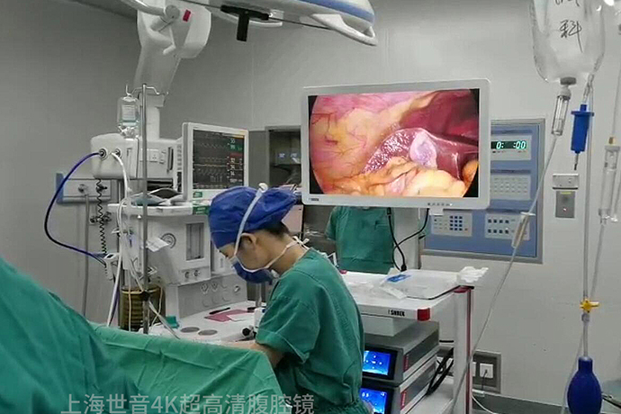
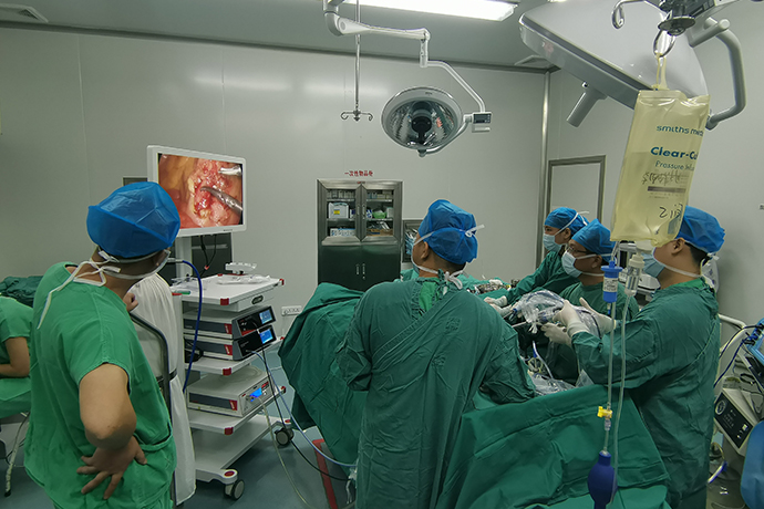
[General Surgery Laparoscopy] 4K Ultra HD Laparoscopic Radical Left Hemicolectomy
Release time: 14 May 2024 Author:Shrek
4K ultra-high definition laparoscopy-assisted radical resection of colon cancer has been accepted by everyone. In recent years, the application of intraluminal anastomosis after laparoscopic left hemicolectomy has gradually increased. This method has the advantages of small incision, mild pain, and quick postoperative recovery, and does not increase the incidence of postoperative complications. It has been gradually accepted by clinicians, and various intraluminal colon anastomosis methods are also emerging. At present, intraluminal colon anastomosis can be performed by manual end-to-end suturing, or by using a stapler to perform end-to-side or end-to-end anastomosis. There is no unified opinion and standard. According to the literature, the mainstream method is side-to-side anastomosis with linear cutting closure device, and the surgeon should flexibly choose and develop an individualized anastomosis method based on personal technical characteristics, specific conditions of the intestinal tract, and the objective economic status of the patient.

Indications
Suitable for splenic flexure, descending colon and sigmoid colon cancer.
Vascular innervation of left colon
1. Inferior mesenteric artery (IMA)
There are many forms of IMA that separate LCA and SA. According to different branch morphology definitions, the proportions reported in the literature are confusing.
From a practical point of view, the author recommends dividing the branch forms of IMA into 3 types:
① LCA and SA form separate types (type I), accounting for about 50%.
②LCA and SA are dry type (II type), accounting for about 40%.
③LCA and the first SA are issued in parallel (type III), accounting for 10%.
2. Middle accessory colic artery
Incidence rate: 4-49.2% (named accessory left colic artery in the literature) Origin point: SMA (cephalad of MCA root)
Course: Covered by the upper edge of the duodenum, passing through the ligament of Treitz, and innervating the splenic flexure and part of the descending colon.
3.Riolan Bow
Riolan Arch: Incidence of anastomotic branches in SMA system and IMA system: 5.5-11.4%.
After the root of the IMA trunk is ligated, the Riolan arch can improve the blood supply of the colon proximal to the anastomosis.
4. Sudeck danger zone
The incidence of missing anastomosis in the marginal arch between the superior rectal artery and the last branch of the SA: 4.7%.
During left hemicolectomy, if the distal bowel remains longer, the IMA should be retained, or the bowel resection in the Sudeck risk zone should be performed simultaneously.
Controversy over the scope of regional lymph node dissection
The extent of lymph node dissection for left colon cancer is currently controversial.
There is still a lack of consensus guidelines specifically focusing on the scope of lymph node dissection for left colon cancer.
There are 3 questions that need clarification:
①D2 or D3?
②What are the D3 lymph nodes, group 223 or group 253?
③Are extra mesangial lymph nodes dissected (such as gastroepiploic arch lymph nodes)?
D2 or D3?
Chinese Colorectal Cancer Diagnosis and Treatment Guidelines (2023 Edition)
T1N0M0 colon cancer + favorable prognostic histologic features: local resection
T2-4N0-2M0: intestinal segmental resection + D3 lymph node dissection
Regional lymph node dissection must include paraintestinal, intermediate, and mesoradicular lymph nodes
If lymph node metastasis beyond the dissection range is suspected, complete resection is recommended
The D3 range of the left colon was not described
Japanese colorectal cancer treatment guidelines (2016 edition, Japanese Colorectal Cancer Association)
cTis can be treated with local resection (D0) or segmental resection (D1)
cSM(T1)N0 patients can undergo D2 surgery
Patients with stage cMP(T2)N0 can undergo D2 or D3 surgery.
For patients with clinical stage 11-111, D3 surgery should be performed
Extra mesangial lymph node dissection
The extra mesangial lymph nodes of the left half of CME mainly refer to the gastroepiploic arch lymph nodes
For example, subpyloric lymph nodes and greater curvature lymph nodes are not routine causes of right colon cancer.
Regional lymph nodes and gastroepiploic arch lymph nodes are not routine causes of left colon cancer.
They drain regional lymph nodes and are “extramesocolic” lymph nodes.
lymph node )
Gastroepiploic arch lymph nodes are lymph nodes within the mesogastric envelope.
It is currently believed that gastroepiploic arch lymph node metastasis in left colon cancer may be related to mesangial
Related to malformed blood vessel communication.
Gastroepiploic arch metastasis occurs in 4-5% of transverse colon cancer and splenic flexure cancer.
Hohenberger proposed the "omental arch principle" during CME: cleaning distance cancer
The omental arch, lymph nodes at the lower edge of the pancreas and the corresponding greater omentum are swollen within 10cm.
Summary 1
Scope of bowel and D3 lymph node resection for left colon cancer:
① IMA-preserving radical surgery for left colon cancer is feasible for splenic flexure cancer, descending colon cancer and descending-descending junction cancer.
, if the distal end of the sigmoid colon is short, radical sigmoid cancer resection with IMA root resection should be performed.
②For splenic flexure cancer, the scope of blood vessel isolation and lymph node dissection should be determined based on the location of the tumor.
: The MCA (dissection 223LN) and LCA (dissection 232LN) are performed for transverse colon cancer near the splenic flexure; 223LN dissection is performed for splenic flexure cancer and the left branch of the MCA is separated, and the LCA is separated (dissection 232LN); the descending colon cancer is dissected near the splenic flexure 253LN And cut off the LCA, cut off the left branch of the MCA (dissection 222LN). In addition, splenic flexure cancer does not require dissection of omental lymph nodes.
The lymph node dissection range for descending colon cancer and cancer at the junction of descending colon cancer and descending colon cancer is: 222LN+253LN.
Lymph node dissection range for sigmoid colon cancer: 253LN.
Summary 2
New central approach: dissection through the lateral approach first, followed by the central approach (below the sacral promontory), which can reduce cross-layer dissection in the left retroperitoneal space.
It is difficult to dissociate the splenic flexure of the colon (especially in obese patients). The three-way outflanking dissociation method mainly through the middle approach is helpful to efficiently dissociate the splenic flexure.
①The method of determining the entry of the upper transverse colon mesocolon into the omental sac according to the T stage (T4 is performed with subarcuate dissociation, non-T4
Perform paracolonic dissection), cut the transverse mesocolon root from inside to outside at the lower edge of the pancreas, and do not simultaneously cut off the third layer of the greater omentum and enter the omental sac (extracapsular method), which is better than cutting the third layer of the greater omentum and entering the omentum at the same time. Capsular (intracapsular method) can significantly reduce the free area of the pancreatic plane, reduce bleeding and pancreatic damage during the separation process (there are reports of postoperative pancreatic leakage due to pancreatic damage), and shorten the freeing time of the splenic flexure.
②Regardless of subarcuate or paracolic dissection, intracapsular or extracapsular method, the path and range of separation at the splenic flexure of the pancreatic tail are the same, that is, the incision is made at the corner of the second and third layers of the greater omentum of the pancreatic tail and runs along the splenic flexure of the pancreatic tail. The space between the mesogastric and transverse mesocolon between layers 3 and 4.

- Recommended news
- 【General Surgery Laparoscopy】Cholecystectomy
- Surgery Steps of Hysteroscopy for Intrauterine Adhesion
- 【4K Basics】4K Ultra HD Endoscope Camera System
- 【General Surgery Laparoscopy】"Two-step stratified method" operation flow of left lateral hepatic lobectomy
- 【General Surgery Laparoscopy】Left Hepatectomy