[General Surgery Laparoscopy] 4K ultra-high definition laparoscopic radical cholecystectomy for the treatment of T3 gallbladder cancer
Release time: 23 Jul 2024 Author:Shrek
Laparoscopic radical cholecystectomy for the treatment of T3 gallbladder cancer is a complex operation that requires surgeons to have highly specialized skills and rich clinical experience.
In laparoscopic radical cholecystectomy, the extent of resection of specific liver segments often needs to be determined based on the TNM stage of the tumor and the extent of liver invasion. With the update of technology and concepts, previous open liver resection procedures can now be safely performed through the laparoscopic approach. During liver lobectomy, in order to achieve radical tumor cure while retaining as much functional liver parenchyma as possible, Ho et al first proposed the concept of laparoscopic limited anatomical hepatectomy (LLAH) in 2013. The core concept is to still retain anatomical liver resection. Compared with traditional anatomical liver resection, there is no significant difference in the long-term oncological effects of LLAH after surgery; because LLAH also retains more functional liver parenchyma, it effectively reduces the postoperative cost. Incidence of liver failure.

Patients benefit from minimally invasive procedures, such as minimal blood loss, less postoperative pain, and shorter hospital stays.
Preoperative preparation
Patient positioning: The patient lies supine on the operating table with legs slightly apart to allow access to the abdomen. General anesthesia: The patient is given general anesthesia to ensure comfort and safety throughout the procedure.
Surgical methods
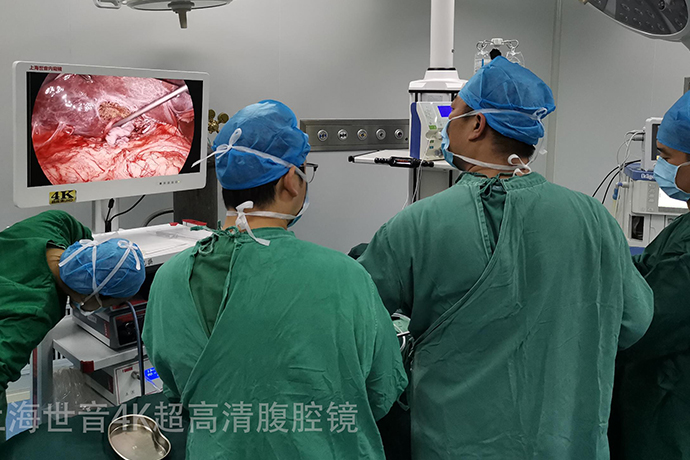
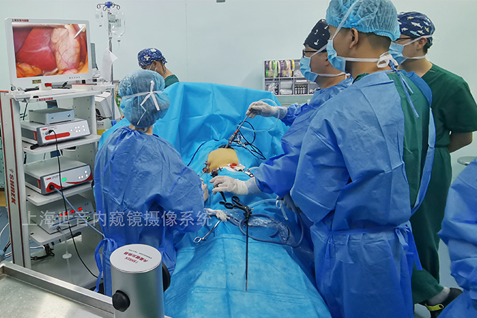
The anesthesia method was tracheal intubation plus intravenous combined general anesthesia, with the patient lying supine on the operating table. The skin was incised 1 cm below the umbilicus and a 10 mm Trocar was inserted to establish pneumoperitoneum, and the pneumoperitoneum pressure was maintained at 10-14 mmHg (1 mmHg=0.133 kPa). Insert a 30° laparoscope to explore the liver and expose its surroundings. Preliminarily confirm the staging of the tumor under the laparoscopic field, explore whether there is peritoneal, liver and distant metastasis, and perform routine pathological examination of the para-aortic lymph nodes (Group 16). , radical surgery for T3 stage gallbladder cancer will be performed after no positive results are found.
The location and scope of liver resection were evaluated, and the remaining Trocars were inserted according to the conventional 5-hole method. The gallbladder triangle is dissected, the cystic artery and cystic duct are ligated and cut, the hepatoduodenal ligament is dissected and a tourniquet is pre-placed (to prevent severe bleeding during the operation), the hepatoduodenal ligament is skeletonized, and lymph nodes are dissected to the metastatic lymph nodes. Next stop is the lymph nodes. Dissect lymph nodes behind the head of the pancreas and on the pancreaticoduodenum as appropriate. Following the principle of en bloc tumor resection, the first porta hepatis was fully dissected, and the right hepatic artery, right branch of portal vein and right hepatic duct were ligated. The perihepatic ligament is severed, and a traditional anatomical right hemihepatectomy or right trilobite resection is performed based on the ischemic line on the liver surface and the anatomical landmarks of the Couinaud liver segment. The right hepatic vein is separated and ligated within the liver parenchyma, and the third stage of extrahepatic treatment with ultrasonic scalpel is performed. Porta hepatitis, patients undergoing right trilobectomy should avoid damaging the left hepatic vein. For patients undergoing limited anatomical hepatectomy, periportal dissection is performed, the sheath of the Glisson system of the right hepatic pedicle is incised, the right hepatic artery, the right branch of the portal vein and the right hepatic duct are separated and then pulled with thin threads without cutting for the time being. According to the Couinaud liver segment Anatomical landmarks were used to resect limited anatomic liver segments in the tumor-bearing portal vein basin. During the separation of liver parenchyma, the involved vessels were ligated and severed with Hem-o-lok clips. Carefully examine and treat liver cross-sections for bleeding and bile leakage.
Insertion of the Laparoscope: A laparoscope is a long, thin, tubular instrument with a camera on the end that is inserted through one of the port incisions. This provides a magnified, high-definition view of the abdominal cavity. Structural identification: The surgeon carefully identifies and evaluates the gallbladder, adjacent liver tissue, lymph nodes, and any suspected areas of tumor involvement. Dissection of Calot's triangle: The cystic duct and cystic artery forming Calot's triangle are dissected and ligated. This allows the gallbladder to be safely separated from the liver.
Discuss
LLAH provides a new way to make radical resection of T3 stage gallbladder cancer more minimally invasive and precise.
For T3 gallbladder cancer diagnosed clearly through preoperative imaging and serological examinations, the relevant professional group guidelines recommend laparotomy. Because gallbladder cancer at stage T3 and above is prone to metastasis to distant sites such as the liver and peritoneum, it is difficult to perform R0 radical resection. Therefore, this type of laparotomy is essentially an exploratory surgery. It is not certain whether the tumor can be cured, and it will inevitably pose a risk to the patient. Cause greater trauma. Experts suggest that laparoscopic exploration with less trauma should be performed first instead of laparotomy. If laparoscopic exploration of gallbladder cancer shows no distant metastasis, given the mature technology and widespread application of laparoscopic precision liver resection in clinical practice, necessary conditions will be created for laparoscopic radical resection of T3 stage gallbladder cancer. In the past, radical resection of T3 stage gallbladder cancer usually required combined anatomical resection of the right hemi-hepatitis or the right three lobes of the liver, with the goal of achieving R0 radical cure of the tumor. However, the remaining liver volume was small, and the incidence of complications such as liver insufficiency after surgery was relatively high. high.With the introduction of the anatomical boundary concept of the tumor-bearing portal vein branch basin centered on the tumor lesion during radical resection of liver cancer, and the application of intra-abdominal ultrasonography, anatomical liver resection is easier to perform. In order to save more functional liver Tissue, LLAH using tumor-bearing intersegmental lesions has been widely used in clinical practice. The development of LLAH provides a new way for radical resection of T3 stage gallbladder cancer to be more minimally invasive and precise. Limited anatomical hepatectomy was first used for liver resection in colorectal cancer liver metastases. Inspired by this, our team applied LLAH in radical resection of T3 stage gallbladder cancer. This study showed that the intraoperative blood loss, postoperative hospitalization time, hospitalization expenses, postoperative complications and incidence of liver dysfunction in the LLAH group were less than those in the traditional group (all P<0.05).
Setting the plane of separation of the tumor-bearing liver parenchyma during radical resection for T3 stage gallbladder cancer is the most critical technical aspect of LLAH.
A limited anatomical liver resection is performed during radical resection of T3 stage gallbladder cancer. The tumor and surrounding tumor-bearing subhepatic segment portal vein branch drainage basin liver segment tissue are removed. The principle of parenchymal preservation is followed to avoid damage to liver function. Therefore, accurate preoperative imaging assessment and setting of the resection plane are key to the application of LLAH in radical resection for T3 stage cholecystocarcinoma. Before surgery, thin-section enhanced CT and MRI three-dimensional visualization technology can clearly display the gallbladder, liver infiltration lesions and involved vascular drainage areas, and plan the best surgical plan. We block the liver pedicle on the diseased side according to the anatomical marks on the liver surface, and then block the ischemic line on the surface of the liver after the liver pedicle, the course of the hepatic veins in the liver parenchyma or the interbranch of the tumor-bearing liver segments, and the retrohepatic inferior vena cava. Cut off the liver parenchyma at a defined plane. However, the Couinaud intersegmental plane of classic anatomy is sometimes not completely consistent with the above-mentioned plane, which is also the reason why there was one R1 resection in each group. Compared with traditional anatomical hepatectomy, limited anatomical hepatectomy requires more strict cross-sectional observation of the reserved liver parenchyma to understand whether there is ischemia or congestion, and whether there is bile leakage, so as to avoid postoperative liver dysfunction.In recent years, indocyanine green fluorescence or GPC3 navigation has gradually been implemented in liver surgery. The identification and confirmation of planes and resection margins in deep liver parenchyma have become more reliable, and the tumor-bearing portal vein branch basin liver The surgical margins of tissue can be accurately and fully displayed to ensure normal blood supply and drainage of the remaining liver after resection. This is expected to be applied in anatomical liver resection for radical resection of T3 cholecyst cancer.
Compared with traditional anatomical liver resection, limited anatomical liver resection is used in laparoscopic radical resection of T3 stage gallbladder cancer. The anatomical section is more detailed and complex, the exposure is relatively difficult, and the operation is more difficult, so the operation time is prolonged to a certain extent. . In addition, laparoscopic radical resection for T3 stage gallbladder cancer also has the problem of intraperitoneal implantation and incisional implantation of gallbladder cancer, which occurred in both the traditional group and the LLAH group in this study. Therefore, it is very important to perform strict tumor-free operations during the operation, avoid gallbladder damage, control the abdominal air pressure and the chimney effect caused by Trocar air leakage, and place all resected specimens in special bags and remove them safely. The advantage of LLAH in this process is that while performing a complete R0 resection, the functional liver structure and volume are preserved as much as possible, so the incidence of postoperative complications and liver dysfunction is low, the patient recovers quickly, and the hospitalization time is shortened. In recent years, maximizing organ protection and achieving the best recovery results with minimal trauma have become the goals of minimally invasive and precise biliary surgery. The application of LLAH in radical resection of gallbladder cancer is a reflection of this concept.

- Recommended news
- 【General Surgery Laparoscopy】Cholecystectomy
- Surgery Steps of Hysteroscopy for Intrauterine Adhesion
- 【4K Basics】4K Ultra HD Endoscope Camera System
- 【General Surgery Laparoscopy】"Two-step stratified method" operation flow of left lateral hepatic lobectomy
- 【General Surgery Laparoscopy】Left Hepatectomy