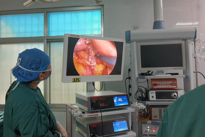
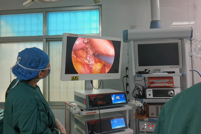
[General Surgery Laparoscopy] 4K Ultra HD Laparoscopic Treatment of Cholecystitis
Release time: 30 Jul 2024 Author:Shrek
What is gallbladder
Gallbladder: with a volume of 40 to 60 ml, it is located in the upper right abdomen of the human body, in the gallbladder fossa behind the liver. It looks like a "fat pear" and is divided into four parts: gallbladder base, gallbladder body, gallbladder neck and cystic duct. It is normally 8 to 12cm long. Width 3~5cm.

Gallbladder function
Secretion of mucus: About 20ml of mucus is secreted every day, which lubricates and protects the mucous membrane.
Concentrated bile: The bile secreted by liver cells (800-1000ml per day) can be concentrated 5-10 times.
Storage of bile: When people are hungry, bile is usually stored in the gallbladder.
Excretion of bile: After the human body eats, the gallbladder begins to contract and discharges the stored bile into the duodenum to aid digestion.
Causes of cholecystitis
Cholecystitis is often caused by bacterial infection secondary to cholestasis caused by gallbladder stones. Irregular diet, overeating, excessive consumption of high-fat and high-cholesterol foods, poor mood, long-term depression, etc. can all cause obstruction of bile excretion and lead to cholestasis.
Classification of cholecystitis
According to the symptoms and course of the disease, it is divided into: acute cholecystitis and chronic cholecystitis.
Acute cholecystitis: sudden onset, short course, inflammation caused by bacterial infection secondary to cystic duct obstruction. After conservative treatment such as fasting, anti-inflammatory, and antispasmodic treatment, the condition can often be relieved; if the inflammation is not effectively controlled, purulent cholecystitis, gallbladder perforation, etc. may ensue. It is currently believed that surgery is still the best treatment for acute cholecystitis.
Chronic (atrophic) cholecystitis: a persistent and recurring inflammatory process in the gallbladder or surrounding tissues caused by a variety of factors. 90% of patients have a history of gallbladder stones and biliary colic; it often manifests as dull pain in the right upper quadrant, poor appetite, Nausea, etc.; B-ultrasound examination shows that the gallbladder has reduced size, thicker wall, and even loses its contractile function; sometimes it is difficult to differentiate from gallbladder cancer; once diagnosed, surgery should be performed as soon as possible.
According to the presence or absence of stones, it is divided into: calculus cholecystitis and acalculous cholecystitis.
Calculous cholecystitis: Cholecystitis associated with stones is called calculus cholecystitis. It often manifests as distended pain and discomfort in the upper abdomen, which is often caused by eating a heavy meal or greasy food. It often attacks acutely at night, accompanied by nausea and vomiting. It is more common in women; it may have mild to moderate fever; if chills or high fever occur, Often indicates gallbladder empyema, perforation, etc.
Acalculous cholecystitis: Cholecystitis without stones is called acalculous cholecystitis. The incidence rate accounts for 5% to 10% of acute cholecystitis. The symptoms are similar to those of acute calculus cholecystitis, and the symptoms are often duplicated by other serious diseases. Covered up, it can easily be misdiagnosed.
Complication
If cholecystitis is not treated promptly and effectively, it can easily lead to complications such as gallbladder empyema, gangrene, or perforation.
Cholecystitis: It is a manifestation of severe gallbladder inflammation, often caused by stones blocking the bile duct, cholestasis in the gallbladder, and bacterial infection. You should cooperate with a doctor for bile culture, use of sensitive antibiotics, puncture drainage, or surgical treatment.
Gallbladder gangrene: It is the most serious type of cholecystitis. It develops from acute cholecystitis and cholecyst empyema. The condition is severe and progresses rapidly. It is more common in middle-aged and elderly patients, accounting for 10% to 40% of acute cholecystitis.
The blood circulation disorder in the gallbladder causes bleeding and gallbladder tissue necrosis; if not treated in time, it can lead to gallbladder perforation, perigallbladder abscess or diffuse peritonitis, etc., with a fatality rate as high as 50%.
Gallbladder perforation: Cholecystitis, gallstones, common bile duct obstruction, etc. can lead to an increase in the pressure in the gallbladder cavity, ischemic necrosis of the gallbladder wall, or the formation of mucosal ulcers, resulting in gallbladder perforation, which is very dangerous; if not treated in time, it can cause septic shock or die.
Treatment method
There are two main surgical treatments for cholecystitis, one is traditional open cholecystectomy, and the other is modern 4K ultra-high definition laparoscopic cholecystectomy. Open cholecystectomy is a traditional surgical method. The doctor will make a large incision in the upper right abdomen and directly remove the inflamed gallbladder. The advantage of this method is that it is direct and thorough, but the disadvantage is that there is greater postoperative pain. , the recovery period is longer.
4K Ultra HD Laparoscopic Cholecystectomy is a minimally invasive surgery. The doctor will make several small incisions in the abdomen and remove the gallbladder through 4K laparoscopy. The advantages of this method are less trauma and less postoperative pain. High fat intake foods to reduce the risk of cholecystitis recurring. Regular follow-up examinations are also required after surgery to evaluate the recovery of the body.
Although the effect of surgical treatment of cholecystitis is usually better, not all patients with cholecystitis are suitable for surgical treatment. For example, for those patients with severe heart or respiratory diseases who cannot bear the risks of surgery, doctors may choose drug treatment. Or try other non-surgical treatments, such as percutaneous gallbladder drainage, a method of draining bile from an inflamed gallbladder through the skin. On the other hand, for patients with recurrent cholecystitis or gallbladder stones, doctors usually recommend surgical treatment, because cholecystitis in these cases is more likely to cause serious complications, and surgical treatment can effectively prevent complications. occur. In general, the treatment of cholecystitis needs to be individualized based on the patient's specific condition. When faced with choosing a treatment plan, patients must consider not only the efficacy of the treatment, but also the risks of the treatment and their own physical condition. Doctors will also consider these factors when formulating treatment plans to ensure that the treatment plan can achieve the purpose of treatment while minimizing harm to the patient.
4K laparoscopic cholecystectomy surgical steps:
1. Routinely disinfect the abdomen and lay down sterile surgical towels. Make an arc-shaped incision along the lower edge of the umbilical fossa, about 10mm long. If the lower abdomen has been operated on, an incision can be made at the upper edge of the umbilical cord to avoid the original surgical scar, and the skin can be incised.
2. Use towel forceps to lift the abdominal wall at the umbilical veress needle, and use a 10mm trocar to puncture. The first puncture has a certain degree of "blindness" and is a more dangerous step in laparoscopy, so be extra careful.
3. Rotate the trocar slowly and insert the needle evenly with force. When entering the abdominal cavity, you will feel a sudden disappearance of resistance. When the closed air valve is opened and gas escapes, the puncture is successful. Connect the pneumoperitoneum machine to maintain constant intra-abdominal pressure.
4. Then insert the laparoscope and puncture each point under the supervision of the laparoscope.
Generally, a 10mm cannula is punctured 2cm below the xiphoid process to prepare discharge coagulation hooks, clip appliers and other instruments; a 5mm cannula is used each 2cm below the costal margin of the right midclavicular line or 2cm below the outer edge of the rectus abdominis and anterior axillary line. Cannula puncture was performed to place the irrigator and gallbladder fixation grasper.
5. At this time, artificial pneumoperitoneum and preparation work have been completed. Due to the creation of pneumoperitoneum and the first trocar puncture, the large internal blood vessels and intestines of the abdominal cavity may be accidentally injured, and it is not easy to find during the operation. Recently, many people make a small opening in the umbilicus, find the peritoneum, and directly insert the trocar into the abdominal cavity to inflate it. After the pneumoperitoneum is successfully created, the surgical operation begins. The division of labor in surgery varies from hospital to hospital. In our case, the surgeon controls the gallbladder fixation forceps and electrocoagulation hook, and is responsible for all operations of the surgery. The first assistant controls the irrigator, which is responsible for flushing, suctioning, and assisting in the exposure of the surgical field; the second assistant controls the irrigator. Laparoscopy keeps the surgical field displayed in the center of the television screen.
6. Use grasping forceps to grasp the neck of the gallbladder or Hartmann's capsule and pull it upward and to the right. It is best to draw the cystic duct perpendicular to the common bile duct to clearly distinguish the two, but be careful not to draw the common bile duct into an angle. Cut the serosa on the cystic duct, bluntly separate the cystic duct and the cystic artery, and distinguish the common bile duct and the common hepatic duct. Since this part is close to the common bile duct, electrocoagulation should be used as little as possible to avoid accidental injury to the common bile duct. Separates the cystic duct up and down. And see clearly the relationship between the cystic duct and common bile duct.
7. Place the titanium clip as close to the gallbladder neck as possible. There should be sufficient distance between the two titanium clips, and the distance between the titanium clip and the common bile duct should be at least 0.5cm.
8. Use scissors to cut between the two titanium clips. Do not use electric cutting or electrocoagulation to prevent damage to the common bile duct due to heat conduction. Then the cystic artery was found posteriorly and medially, and a titanium clip was placed to cut it. After cutting off the cystic duct, do not pull hard to avoid severing the cystic artery, and pay attention to the posterior branch vessels of the gallbladder. The gallbladder is carefully dissected, and bleeding is stopped by electrocoagulation or titanium clips.
9. Clamp the gallbladder neck and pull it upward, and carefully peel it along the gallbladder wall. The assistant should assist in pulling so that there is a certain tension between the gallbladder and liver bed. Peel off the gallbladder completely and place it on the upper right side of the liver. Use electrocoagulation to stop bleeding in the liver bed, rinse it carefully with normal saline, and check for bleeding and bile leakage (put a piece of gauze on the liver porta and remove it to check for bile staining).
10. After draining the intra-abdominal fluid, transfer the laparoscope to the subxiphoid cannula and make way for the umbilical incision so that the gallbladder containing stones >1cm can be removed from the umbilical incision, which is relatively loose in structure and easy to expand. If the stone is small, it can also be removed from the puncture hole under the xiphoid process.
11. Send the toothed grasping forceps from the umbilical trocar into the abdominal cavity, grasp the stump of the cystic duct under supervision, slowly drag the gallbladder into the trocar sheath, and pull it out together with the trocar sheath. When grasping the gallbladder, be careful to place the gallbladder on the liver to avoid accidentally injuring the intestines with the sharp forceps. If the stone is large or the gallbladder tension is high, do not pull it out with force to avoid gallbladder rupture and leakage of stones and bile into the abdominal cavity.
12. You can use vascular forceps to enlarge the incision and take it out. You can also use a dilator to expand the incision to 2.0cm. If the stone is too large, you can extend the incision. If bile leaks into the abdominal cavity, apply wet gauze through the umbilical incision to suck out the bile. When the stones are too big to be removed from the incision, you can also open the gallbladder first, use a suction device to suck out the bile in the gallbladder, crush the stones and remove them one by one. If any stones are found falling into the abdominal cavity, they should be removed.
13. After checking that there is no accumulation of blood or fluid in the abdominal cavity, pull out the laparoscope, open the valve of the cannula to drain the carbon dioxide gas in the abdominal cavity, and then pull out the cannula. Use a thin thread to suture the fascial layer for 1 or 2 stitches at the incision where the 10mm cannula is placed, and close each incision with sterile tape.
Cholecystitis is a disease that can be effectively treated with surgery, but this does not mean that we can ignore its prevention. Eating a healthy diet, controlling your weight, and avoiding excessive drinking are all important measures to prevent cholecystitis. Only if we pay attention to both treatment and prevention can we truly control cholecystitis and protect our health.

- Recommended news
- 【General Surgery Laparoscopy】Cholecystectomy
- Surgery Steps of Hysteroscopy for Intrauterine Adhesion
- 【4K Basics】4K Ultra HD Endoscope Camera System
- 【General Surgery Laparoscopy】"Two-step stratified method" operation flow of left lateral hepatic lobectomy
- 【General Surgery Laparoscopy】Left Hepatectomy