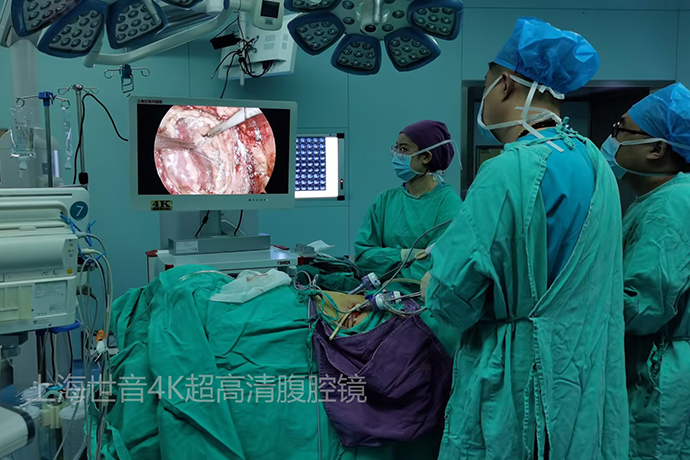
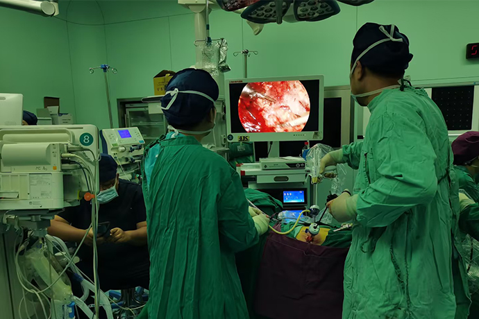
[General Surgery Laparoscopy] Radical right nephrectomy under 4K ultra-high definition laparoscopy
Release time: 20 Aug 2024 Author:Shrek
In the past 20 years, the incidence rate of kidney cancer in my country has been increasing at an astonishing rate of 6.5% per year, ranking second among urological cancer diseases, and has become the "main killer" of deaths from malignant tumor diseases.
Our human kidneys are composed of many nephrons, and these nephrons do not work at the same time. On the one hand, this "rotation system" protects our kidneys from being too "tired", but it also causes There will be certain negative effects. That is why many people may not even notice when their kidneys have problems. Over time, the condition will worsen, and it may even be found at an advanced stage, making it difficult to cure.

Therefore, in daily life, if people discover their own problems, they must go to the hospital for examination to protect their health.
Treatment options
In general, kidney cancer is most commonly treated by surgical removal.
Surgery is the main treatment for kidney cancer that has not spread outside the kidneys. Depending on the type of kidney cancer, the stage and grade of the cancer, and your general health, you may do one of the following:
1. Removal of the entire kidney (radical nephrectomy)
This is the most common surgery for large tumors. The entire affected kidney, a small portion of the ureter, and surrounding fatty tissue are removed. The adrenal gland and nearby lymph nodes may also be removed. Sometimes, kidney cancer may have spread into the renal veins or even into the vena cava, which is a part of the body primarily close to the spine. Even if the cancer is in the vena cava, sometimes all the cancer can be removed in one surgery.
2. Removal of part of the kidney (partial nephrectomy)
This is sometimes an option for tumors that are limited to the kidneys, and is particularly useful for people with preexisting kidney disease, kidney cancer, or those with only one working kidney. Only the cancer and a small part of the kidney are removed, which means the kidney's function is preserved. Partial nephrectomy is more difficult to perform than radical nephrectomy and may depend on the location of the tumor.
If the entire kidney or part of the kidney is removed, the remaining kidney usually does the work of two kidneys.
How the surgery is done
If you have kidney cancer surgery, it will be done in a hospital under general anesthesia.
Whether all or part of the kidney is removed (radical or partial nephrectomy), different surgical approaches are available, each with advantages in specific circumstances.
laparoscopic surgery
Laparoscopic surgery is sometimes called keyhole or minimally invasive surgery. The surgeon will make several small incisions and insert a tiny instrument with a light and camera (laparoscope) into one of the incisions. The laparoscope takes a picture of your body and forwards it to a TV screen. The surgeon inserts the tool into the other incisions. and perform surgery using on-screen images for guidance.
Standard laparoscopic and robot-assisted surgeries generally mean shorter hospital stays, less pain and faster recovery times compared to open surgeries. However, in some cases, open surgery may be a better option.
Case
The patient was initially diagnosed with right renal cancer and underwent retroperitoneal right radical nephrectomy.
Contrast-enhanced CT of the urinary tract showed space occupying the lower pole of the right kidney, which was significantly enhanced after contrast enhancement. The diagnosis was considered to be right renal cancer.
There are small branches of the renal arteries at the lower pole of the kidney that supply the tumor. Dissect along the dorsal layer of the psoas major muscle toward the deep side to find the exposed inferior vena cava. Often the inferior vena cava can be isolated bluntly at the dorsal level alone. However, the patient in this case had severe adhesions, and the fat on the surface of the inferior vena cava was sharply incised during the operation. Dissociate along the surface of the inferior vena cava and cut the inferior vena cava vascular sheath (which is a relatively avascular zone). When incision, pay attention to the technique of the ultrasonic scalpel blade, pull it upward and then perform the incision to avoid the blade pointing directly at the inferior vena cava and causing perforation and bleeding. It was dissected along the level of the inferior vena cava, from the foot to the head, and encountered the main trunk of the right renal artery.
Use the Hem-o-lok vascular clamp to clamp and cut the right renal artery, continue to dissociate cephalad along the surface of the inferior vena cava, and cut the connective tissue outside the inferior vena cava sheath. Turn the direction of dissection to the foot side, and find the thickened gonad vein and the small branches of the arteries supplying the tumor at the lower pole of the kidney. Use vascular clips to clamp and cut off the small arteries and thickened gonadal veins respectively. The right ureter below the lower pole of the kidney was freed, and the Hem-o-lok vascular clamp was used to clamp the ureter. The right ureter was cut with scissors, and the blood spurting from the ureteral nutrient artery was visible. For blood spurting from the ureteral nutrient artery, the ultrasonic scalpel is used to coagulate and sever the nutrient artery at the same time. When dissecting the ventral level (the location of the lower pole of the kidney), it can be seen that there are abundant thickened collateral veins in the fat sac of the lower pole of the kidney. After the tortuous vein bleeds, the left hand is helping to cover the lower pole of the right kidney. The right hand ultrasonic knife quickly clamps the bleeding vein. The bleeding site is determined as soon as possible and the bleeding vein is clamped in time within the "golden three seconds" of bleeding.The right hand remains motionless after clamping the vein, and the left hand curved forceps moves and takes over from the right hand to clamp the bleeding vein. Keep the curved forceps in your left hand to clamp the bleeding vein, free the ultrasonic scalpel in your right hand, and use electrocoagulation to close and cut off the tortuous bleeding vein. After coagulation, a vascular clamp is used to clamp the tortuous bleeding vein at the proximal renal end, and the bleeding point is coagulated with bipolar electrocoagulation. Complete hemostasis. After free crossing over the suprarenal pole, care should be taken to preserve the right adrenal gland, and the fat sac of the suprarenal pole should be incised to leave the right adrenal gland in the fat. In this operation, the inferior vena cava was fully freed, and when the adrenal gland was separated, the inferior vena cava deep behind it could be viewed directly, thereby avoiding the ultrasonic scalpel blade from penetrating and damaging the deep inferior vena cava.
In this operation, it was difficult to cut off the right renal vein freely due to the huge tumor and small space. The sequence of steps in the surgical strategy is: free the dorsal level of the kidney → cut off the right renal artery, gonadal vein, and ureter → free the ventral level of the kidney → free the upper pole of the kidney to preserve the adrenal gland → cut off the right renal vein. The right renal vein is the final step of the operation, and can be freely exposed from three levels: dorsal level, ventral level, and upper pole level. The process of the right renal vein was freely exposed from the dorsal level, and it was seen that there was bleeding caused by small branches at the proximal end of the right renal vein. Bipolar electrocoagulation was used to close the bleeding point.The right renal vein was freed and exposed from the ventral level of the kidney, and a right-angle forceps was used to free and expose the right renal vein. From the level of the upper pole of the kidney, press down on the kidney to free and expose the right renal vein. Finally, select the best level (dorsal level) to clamp the Hem-o-lok vascular clip and cut off the right renal vein.

- Recommended news
- 【General Surgery Laparoscopy】Cholecystectomy
- Surgery Steps of Hysteroscopy for Intrauterine Adhesion
- 【4K Basics】4K Ultra HD Endoscope Camera System
- 【General Surgery Laparoscopy】"Two-step stratified method" operation flow of left lateral hepatic lobectomy
- 【General Surgery Laparoscopy】Left Hepatectomy