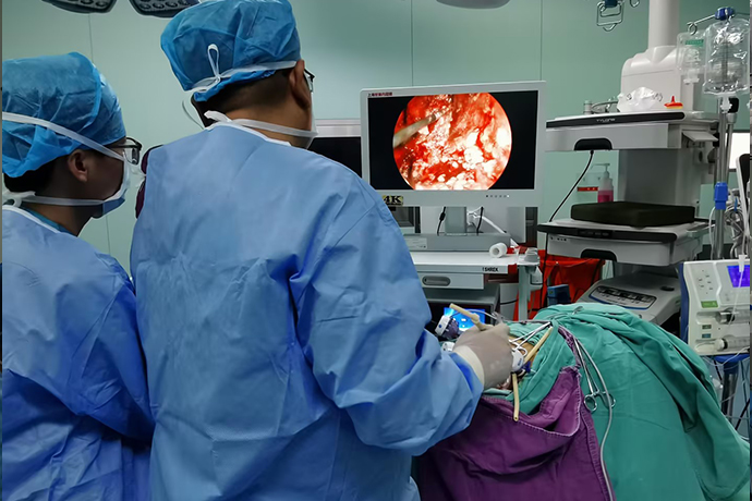
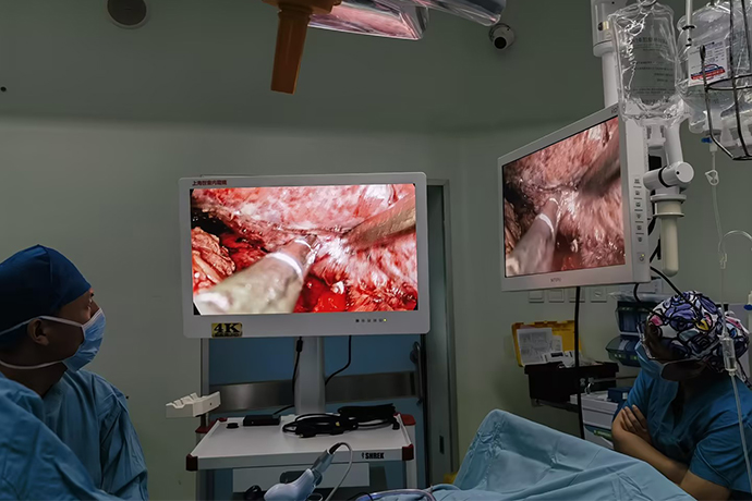
[General Surgery Laparoscopy] 4K ultra-high definition laparoscopic pyeloplasty
Release time: 13 Aug 2024 Author:Shrek
Pyelvesteroplasty is a surgery used to treat kidney disease. It is an operation that establishes the funnel-shaped connection between the renal pelvis and the ureter through suturing to restore physiological, myogenic ureteral and renal pelvic peristalsis to protect kidney function.

Surgical methods include open surgery and laparoscopic surgery. Now as minimally invasive technology matures, especially now that advanced medical device technology is taking shape and being put into clinical use, 4k ultra-high definition laparoscopic pyeloplasty has become the main surgical method.
Indications
For example, obstruction of the ureteral junction caused by abnormal blood vessels coexists with other obstructive lesions in the wall and lumen, such as muscle fiber dysplasia, organic stenosis of the ureter caused by long-term vascular compression, or the presence of wrinkles or membranes in the lumen, etc., when dissociated After the junction of the blood vessels and the ureter, the kidney still cannot empty, and the lumen cannot be filled and expanded in time. Check that the narrow ureteral section is thin and hard. If the peristaltic wave conduction of this section of the ureter is poor, re-anastomosis of the kidney and ureter should be performed. technique. Although there is no vascular obstruction, there are similar organic lesions at the junction of the ductus duct and it is not suitable to undergo other ureteroplasty operations. This is also an indication for nephroureteroplasty. The technical operation of this operation is relatively simple and the effect is good.
Clinical manifestations
Lumbar masses can be seen in children, and intermittent low back pain can be seen in children or adults, sometimes with nausea, vomiting, hematuria, sometimes with upper urinary tract infection, and sometimes with microscopic hematuria or pyuria.
Anesthesia and positioning
Lying on the healthy side, waist high, head and feet low, in an arched bridge position.
Surgery case
1. Free prerenal plane
2. Free the junction between renal pelvis and ureter
3. Remove the stenotic segment of the pelvis-ureter junction
4. Suspension of renal pelvic wall
5. Determine the lowest point of the renal pelvis and place a needle and thread
6. Dissect the ureteral wall longitudinally
7.Posterior wall anastomosis
8. Indwelling ureteral double J tube
9. Suturing the anterior wall of the anastomosis
10. Suture the renal pelvis tear
11. Close the prerenal fascia
Post-operative care
After surgery, the patient should adopt a semi-recumbent position, which can raise the waist and prevent the reflux of urine. If physical strength allows, he should go to the ground to move, which can promote recovery. He should also drink more water, urinate frequently, and never hold urine. , otherwise it will easily cause urinary system infection. Patients should also pay attention to daily care after being discharged from the hospital, and go to the hospital for regular check-ups, such as B-ultrasound and CT examinations.

- Recommended news
- 【General Surgery Laparoscopy】Cholecystectomy
- Surgery Steps of Hysteroscopy for Intrauterine Adhesion
- 【4K Basics】4K Ultra HD Endoscope Camera System
- 【General Surgery Laparoscopy】"Two-step stratified method" operation flow of left lateral hepatic lobectomy
- 【General Surgery Laparoscopy】Left Hepatectomy