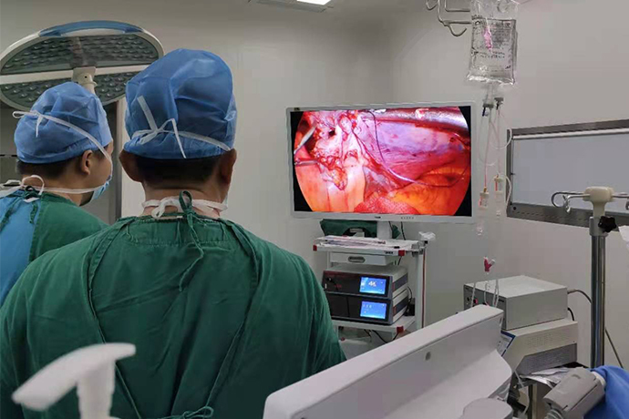
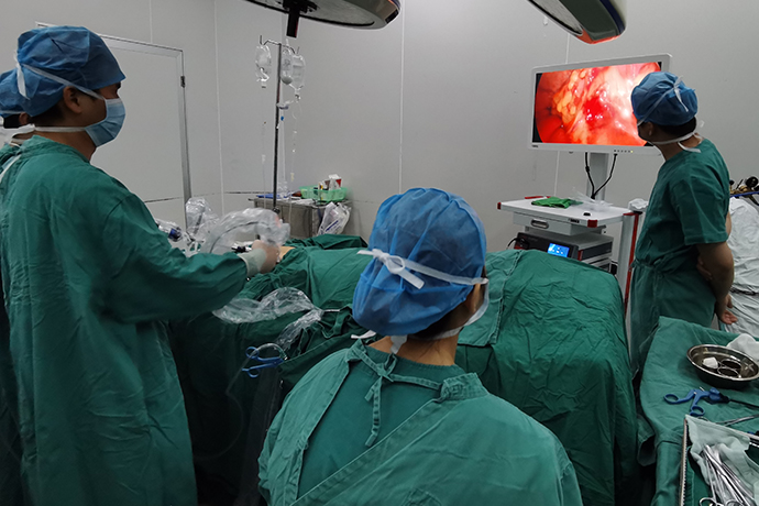
[General Surgery Laparoscopy] 4K Ultra HD Laparoscopic Right Hemicolectomy
Release time: 21 May 2024 Author:Shrek
Colon cancer is a common malignant tumor of the digestive tract that occurs in the colon. It mostly occurs at the junction of the rectum and the sigmoid colon. The incidence rate is highest in the 40- to 50-year-old age group, with a male-to-female ratio of 2 to 3:1. The incidence rate ranks third among gastrointestinal tumors. Colon cancer mainly includes adenocarcinoma, mucinous adenocarcinoma, and undifferentiated carcinoma. The general shape is polypoid, ulcer-like, etc. Colon cancer can develop circularly along the intestinal wall, spread up and down along the longitudinal diameter of the intestinal tube, or infiltrate deep into the intestinal wall. In addition to metastasis and local invasion through lymphatic vessels and blood flow, it can also be planted in the abdominal cavity or spread along sutures and incision surfaces. . Patients with chronic colitis, patients with colon polyps, and obese men are susceptible groups.
With the advancement of medical technology, minimally invasive surgery is now widely used in the treatment of colon cancer. This article combines information on the Internet to sort out laparoscopic right hemicolectomy.

Indications:
1. Cecum cancer, ascending colon cancer and colon and hepatic flexure cancer.
2. Appendiceal adenocarcinoma that invades the cecum or has lymph node metastasis.
Contraindications:
1. For advanced colon cancer, it is estimated that it is difficult to remove all metastatic lymph nodes.
2. Those with cardiopulmonary disease who cannot undergo tracheal intubation and general anesthesia.
3. Those with a history of upper abdominal surgery and extensive adhesions in the upper abdomen.
Anesthesia method
1. Continuous epidural anesthesia or general anesthesia.
Surgical steps:
1. Establish pneumoperitoneum and explore the abdominal cavity
2. Handle blood vessels: anatomical landmark---superior mesenteric vessel
Treat blood vessels
GTH: gastrocolic trunk (Henle’s trunk) RGEV: right gastroepiploic vein MCV: middle colonic vein RCV: right colic vein SRCV: superior right colic vein (accessory right colic vein) SMV: superior mesenteric vein
ASPDV: anterior inferior pancreaticoduodenal vein
Treat blood vessels
The ileocolic arteries and veins were cut off by root ligation.
Treat blood vessels
The right colic artery was severed by root ligation.
The right colic vein was severed by root ligation.
It was separated to Henle’s trunk, the surrounding lymph nodes were cleaned, the right colic vein and right gastroepiploic vein were dissected out, and the right colic vein root was ligated and severed.
The mid-colonic blood vessels were processed, and their right branches were separated along the right edge of the mid-colonic artery and vein and ligated and severed.
The greater omentum and right gastrocolic ligament were mobilized.
The hepatic flexure of the colon was freed and the lateral peritoneum was separated.
The space between Toldt and Gerota's fascia was entered for dissection, the right mesocolon and the posterior wall of the intestinal tube were peeled off, and the right colon was completely freed.
The left colon was resected and ileo-transverse colon anastomosis was performed.
Advantages of Laparoscopic Right Hemicolectomy
The magnifying effect of the laparoscope makes the surgical field clear, making it easier to perform sharp separation in the tissue gap. The operation is more precise, and important blood vessels can be clearly exposed, reducing damage and bleeding. During the operation, the ultrasonic scalpel is used to precisely cut and stop bleeding, reducing the amount of bleeding and the incision is small. Less invasive and beautiful.
Gastrointestinal function recovers quickly, which is beneficial to patient recovery and early adjuvant treatment to reduce post-operative stress response.
Postoperative complications
1. Anastomotic leakage.
2. Anastomotic stenosis.
Postoperative care
1. Continue gastrointestinal decompression until intestinal peristalsis is restored and the anus is exhausted, then it can be removed. Intravenous fluids must be administered during decompression.
2. Continue to use antibiotics.
3. From the 5th day after surgery, take liquid paraffin orally every night.
4. Closely observe the wound for bleeding.
5. Strengthen nutrition and management.
Precautions
Follow-up visits on time after discharge. If you suddenly feel unwell after discharge, such as abdominal pain, abdominal pain, jaundice, etc., seek medical advice promptly. You can do some outdoor activities with a small amount of exercise, such as walking after meals, playing chess, Tai Chi and other outdoor activities. To prevent colds, avoid smoking and drinking, and try to avoid going to crowded public places.
Postoperative diet
You can drink a small amount of water on the second day after surgery, liquid food on the third day, semi-liquid food on the fifth day, and then gradually change to soft food according to the situation. Eat high-calorie, high-protein, high-vitamin, low-fat, easy-to-digest foods, and eat less animal fat, animal offal, fried and spicy foods. Eating method: Eat small meals frequently, 5 to 6 meals per day, eat regularly, pay attention to food matching, and have reasonable nutrition.

- Recommended news
- 【General Surgery Laparoscopy】Cholecystectomy
- Surgery Steps of Hysteroscopy for Intrauterine Adhesion
- 【4K Basics】4K Ultra HD Endoscope Camera System
- 【General Surgery Laparoscopy】"Two-step stratified method" operation flow of left lateral hepatic lobectomy
- 【General Surgery Laparoscopy】Left Hepatectomy