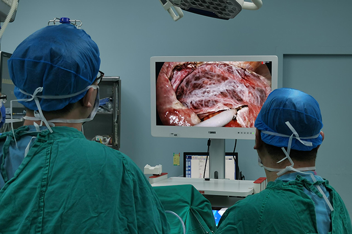
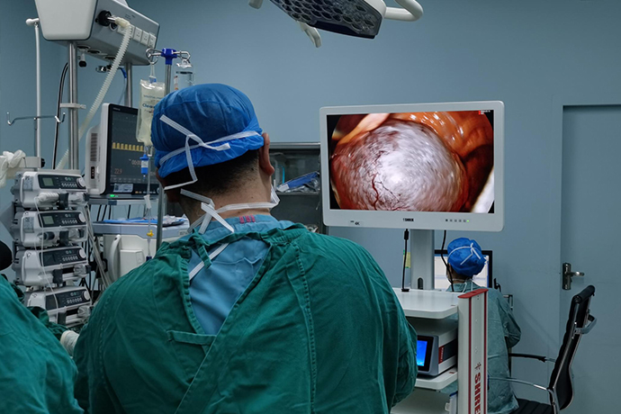
[General Surgery Laparoscopy] 4K Ultra HD Laparoscopic Splenectomy
Release time: 28 May 2024 Author:Shrek
Splenectomy is widely used in diseases such as splenic rupture, wandering spleen (ectopic spleen), local spleen infection or tumor, cyst, intrahepatic portal hypertension combined with hypersplenism and other congestive splenomegaly. The spleen is the largest peripheral lymphoid organ in the human body. It can produce a variety of immunoactive cytokines. It is the main organ of the body for blood storage, hematopoiesis, blood filtration, and blood destruction. It has important immune regulation, anti-infection, anti-tumor, endocrine and production functions. Properdin and pro-phagocytic peptides. Based on the current understanding of spleen function and the increased susceptibility of patients to infection after splenectomy, it is the consensus of surgeons around the world to perform spleen-preserving surgery as much as possible when conditions and disease permit. That is to say, "saving life is the first priority, preserving the spleen is the second priority. The younger you are, the priority is to preserve the spleen."

Case sharing
Basic information: The patient, female, 60 years old, was admitted to the hospital with the main complaint of "diagnosed schistosomiasis cirrhosis with splenomegaly and hypersplenism for more than ten years." She was treated with long-term oral medications and required surgical treatment.
Physical examination on admission: face with chronic liver cirrhosis, mild anemia, skin and sclera not yellow, flat abdomen, no obvious abdominal varicose veins, soft palpation, spleen 3 transverse fingers under the ribs, liver not palpable under the ribs, no shifting dullness , bowel sounds are normal. There is no edema in the limbs.
Auxiliary examination: Routine blood test showed reduction of three systems and mild anemia. Eight viral tests, five tumor markers, and biochemical and coagulation functions were normal.
Gastroscopy: It showed mild esophageal and gastric varices and portal hypertensive gastritis.
CT: It indicates liver cirrhosis, liver lobe imbalance, and splenomegaly.
Surgical procedure
1. Explore the abdominal cavity, open the omental sac to the splenic hilum, and pre-treat some short gastric blood vessels.
2. Separate the pancreaticogastric ligament; suspend the greater curvature of the gastric body.
3. Separate the adhesive omentum tissue in the splenic hilum area to expose the splenic hilum.
4. Separate the adhesions of the lower pole of the spleen and the splenocolic ligament.
5. Isolate and process the lower pole blood vessels of the spleen.
6. Hold up the lower pole of the spleen and separate the lower part of the splenorenal ligament and the retrosplenic hilum space.
7. Isolate the splenic artery at the upper edge of the pancreas and ligate it with No. 7 silk suture.
8. Open the retroperitoneum on the inside of the upper spleen capsule above the tail of the pancreas, gradually separate the spleen and stomach fundus space upward along the direction of the stomach fundus, and deal with the remaining short gastric blood vessels.
9. Lift the upper pole of the spleen, separate the splenophrenic ligament and the upper part of the splenorenal ligament, and join the lower part.
10. Use a golden finger to penetrate the tunnel behind the splenic hilum and tie the splenic pedicle with No. 7 silk suture.
11. Then use gold fingers to penetrate the posterior tunnel of the splenic pedicle, and use Endo-GIA blue nails or white nails to close and separate the splenic pedicle.
12. Check the splenic pedicle stump and strengthen it with titanium clips.
13. Separate the residual splenorenal ligament.
14. Bag the specimen and remove the specimen through the enlarged umbilical hole.
15. Re-enter the microscope to check for bleeding from the wound, whether there is any damage to the gastric wall, and whether the closed stump is reliable.
16. Place drainage tubes in the splenic fossa and splenic pedicle section.
17. Remove the gastric suspension.
18. Remove pneumoperitoneum and suture the incision.
Postoperative pathology
Gross: 1 spleen specimen, approximately 15.0 cm × 8.2 cm × 3.0 cm in size. Part of the spleen tissue was severed. The cross-section was bean paste red. No hematoma was visible in the parenchyma. No obvious gray-white mass was found in multiple cross-sections.
Pathological diagnosis: chronic congestive splenomegaly.
Postoperative precautions
1. Observe whether there is internal bleeding, and routinely measure changes in blood pressure, pulse and hemoglobin. Observe the condition of the drainage tube in the splenic fossa under the diaphragm. If there is a tendency of internal bleeding, timely blood transfusion and fluid replenishment should be carried out. If the bleeding is sustained, reoperation should be considered to stop the bleeding.
2. Splenectomy causes great stimulation to the abdominal organs (especially the stomach), so a gastrointestinal decompression tube should be installed to prevent gastric dilatation after surgery. You should resume eating 2 to 3 days after surgery.
3. Many patients who undergo splenectomy have poor liver function. Vitamins, glucose, etc. should be fully supplemented after surgery. If hepatic coma is suspected, corresponding preventive and therapeutic measures should be taken promptly.
4. Pay attention to changes in kidney function and urine output, and be alert to the occurrence of hepatorenal syndrome.
5. Antibiotics are routinely used after surgery to prevent and treat systemic and subphrenic infections.
6. Measure the platelet count promptly. If it rises rapidly to above 50×109/L, splenic vein thrombosis may occur. If severe abdominal pain and bloody stools occur again, it indicates that the thrombus has spread to the superior mesenteric vein, and antibiotics must be used promptly. Coagulation treatment and surgery if necessary.

- Recommended news
- 【General Surgery Laparoscopy】Cholecystectomy
- Surgery Steps of Hysteroscopy for Intrauterine Adhesion
- 【4K Basics】4K Ultra HD Endoscope Camera System
- 【General Surgery Laparoscopy】"Two-step stratified method" operation flow of left lateral hepatic lobectomy
- 【General Surgery Laparoscopy】Left Hepatectomy