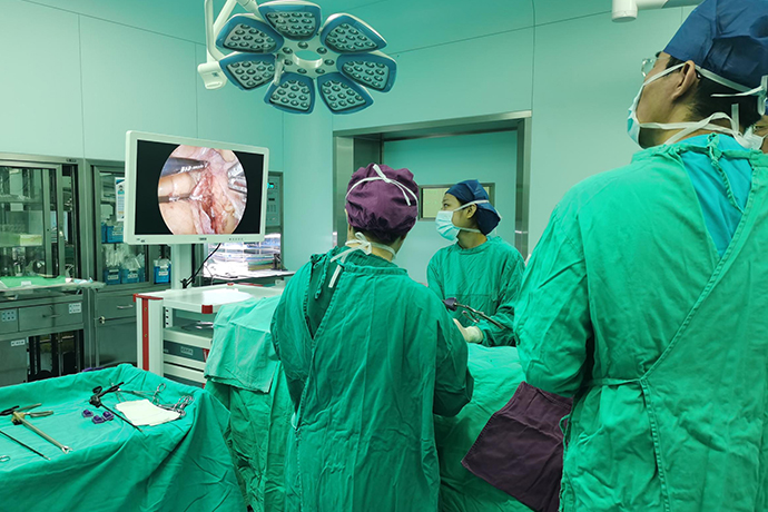
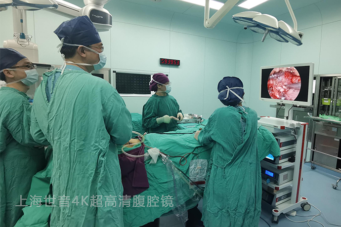
[General Surgery Laparoscopy] Radical cystectomy under 4K ultra-high definition laparoscopy
Release time: 11 Jun 2024 Author:Shrek
The bladder is a muscular sac-like organ that stores urine. When empty, it is in the shape of a triangular pyramid. The top is tapered and faces forward and upward, which is called the bladder apex. It is connected to the umbilicus by the median umbilical ligament. The base is triangular and faces backward and downward, called the bladder base. , most of the area between the tip and the base is called the body of the bladder, the lower part of the bladder becomes thinner and is called the bladder neck, and the area between the left and right ureteral orifices and the internal urethral orifice in men is called the bladder triangle.

1. Total cystectomy removes the bladder, prostate and seminal vesicles in men, and removes the bladder and urethra in women. Radical total cystectomy is the en bloc removal of the bladder, prostate, seminal vesicles, pelvic peritoneum, pelvic side walls and surrounding tissues of blood vessels (including lymph nodes and lymphatic vessels). 2. In women, it also includes the broad ligament, uterus, cervix and part of the vagina.
Bladder cancer is a common tumor of the urinary system, with a male to female ratio of approximately 3:1.
Radical cystectomy is the gold standard treatment for muscle-invasive bladder cancer.
Evidence-based medicine shows that minimally invasive treatments such as laparoscopy can achieve the same treatment results as open surgery.
In 1954, SAYEGH et al. first reported radical cystectomy for the treatment of advanced bladder cancer.
In 1988, Cuschieri reported for the first time a successful laparoscopic cholecystectomy in experimental animals. In 1992, Para et al first introduced laparoscopic cystectomy technology.
In 2000, Gill et al reported completely laparoscopic radical cystectomy plus urinary diversion.
So far, with the accumulation of surgical experience and the development of medical equipment and instruments, laparoscopic or robot-assisted laparoscopic radical cystectomy has been adopted.
Indications:
1. T2-T4a, N0-x, M0 invasive bladder cancer
2. High-risk non-muscle-invasive bladder cancer T1G3 (high grade)
3. Tis that is ineffective in Bacillus Calmette-Guérin (BCG) treatment
4. Recurrent non-muscle invasive bladder malignant tumors
5. Extensive papillary lesions and non-urothelial bladder cancer that cannot be controlled by transurethral resection and bladder instillation.
Laparoscopic radical cystectomy (men)
Laparoscopic bladder resection, step by step, don’t panic
Position your head with your head down and your legs up, with the intestines inserted into the abdominal cavity as much as possible
Free the left and right ureters to protect the blood supply and not mess around
Umbilical artery is easy to find and early treatment will reduce bleeding.
Lift the seminal vesicles and vas deferens, and Di's fascia can be easily opened.
Separate only when fat is seen to ensure there is no problem in the rectum
Open the pelvic fascia and tie the DVC with "8" sutures
Lateral ligaments, slow processing, little secrets hidden inside
Please save your sexual nerves in exchange for a good reputation as a doctor.
The urethra is first closed and then severed, and the bladder is placed in the abdominal cavity
Exposed pelvic lymphatic clearance, vascular neuroskeletalization
Total bladder resection is very complicated, and it doesn’t matter if the steps are clear.
Laparoscopic radical cystectomy (women)
Female total bladder, different forms
Either finish the pot in one go, or move forward
The suspensory ligament of the ovary is cut first, and then the vaginal vault is opened.
Lateral ligaments, slow processing, there are also little secrets inside
Go around to the front and cut off the urethra, removing the bladder and uterus completely
If only the bladder is cut, the reproductive blood supply should not be damaged
There is a gap between the bladder and uterus, and it is judged that the water sac is pulled at the neck orifice
In order to achieve good urinary control in situ, the urethra should be kept long enough
The specimen can be taken out from the vagina and a bladder can be made in the abdominal cavity
Familiar anatomy and calmness make it easier for women to undergo surgery
Brief surgical steps
Material preparation:
Abdominal bag, clothing 4, endoscopic bag, thermos cup, 11#23# blade, knife handle, ligation clip, No. 1, 4, 7 silk thread, endoscopic cover, cable 4-0, chest drainage tube, various models Suture needle, specimen bag, paraffin oil, ultrasonic scalpel, gastrointestinal chukka, electrosurgical scalpel, a set of suction devices, No. 16 disposable urinary catheter, drainage bag, urinary bipolar (straight blue handle), urinary suction device, intracavitary slit device assembly.
Puncture placement position:
Lens hole A: 10mm puncture device on the upper edge of the umbilicus. Operating hole B: Max point 12mm puncture device. Operating hole C: Anti-McFarland point 12mm puncture needle. Operating hole D: Insert a 12mm puncture device 3cm medial to the left anterior superior iliac spine.
Free the ureter and clean both sides of the pelvic lymph nodes: open the retroperitoneum where the ureter crosses the iliac blood vessels, free the ureter from the inside of the external iliac artery to the entrance of the bladder, use non-invasive grasping forceps and ultrasonic scalpel to free and remove the left and right internal and external iliac lymph nodes and obturator lymph nodes in sequence. Remove with stone pliers.
The two side walls and bottom wall of the bladder are freed, and the bilateral vas deferens and seminal vesicles are freed. The vas deferens is severed, the bottom of the bladder is separated into the Dischmann's space, and the rectum is pushed downward until the tip of the prostate.
Free the lateral bladder ligaments and prostatic ligaments, separate the urethra, and remove the prostate: use ultrasonic knife to separate, incise the peritoneum from the top of the bladder to the retropubic and preprostatic space, open the pelvic floor fascia, free the prostate tip, and perform 2-0 barbs The dorsal vein complex of the penis is sutured, the apex of the prostate and the urethra are severed, and the entire bladder is completely removed.
Specimen removal: Incision below the umbilical cord, longitudinal incision into each layer of the abdominal wall, into the abdominal cavity, and removal of the entire resected bladder. After taking out the specimen, rinse it regularly with warm sterilized water, hold it in the head-high and soles position for 5 minutes, and then aspirate it clean (first, to rinse the pelvic floor, retain the perfusion fluid for a few minutes, and second, to slow down the blood circulation of the head) .
Ureterabdominal wall stoma: free the sigmoid mesocolon, pass the left ureter from behind the sigmoid colon, insert F7 single J tubes into both ureters, and fix the stoma with 0/5 Wei Qiao suture.
Count the items and suture the incision: place a latex tube in the pelvis, count the instruments and gauze and suture the incision.
Postoperative care and recovery: The patient’s vital signs need to be closely observed after the operation, and attention should be paid to the nature and amount of drainage fluid. After the patient resumes eating, nutritional support is required to promote recovery. At the same time, early rehabilitation training should be carried out to improve the patient's quality of life.

- Recommended news
- 【General Surgery Laparoscopy】Cholecystectomy
- Surgery Steps of Hysteroscopy for Intrauterine Adhesion
- 【4K Basics】4K Ultra HD Endoscope Camera System
- 【General Surgery Laparoscopy】"Two-step stratified method" operation flow of left lateral hepatic lobectomy
- 【General Surgery Laparoscopy】Left Hepatectomy