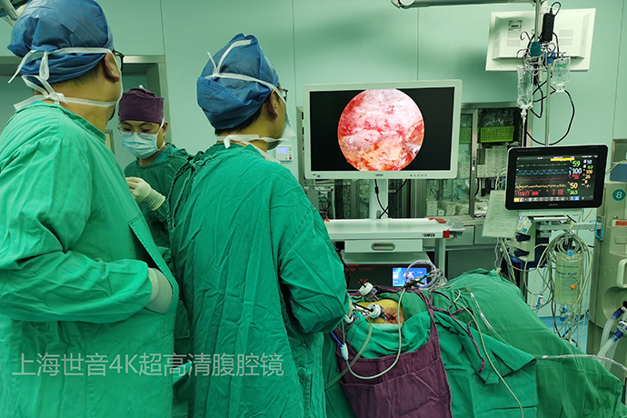
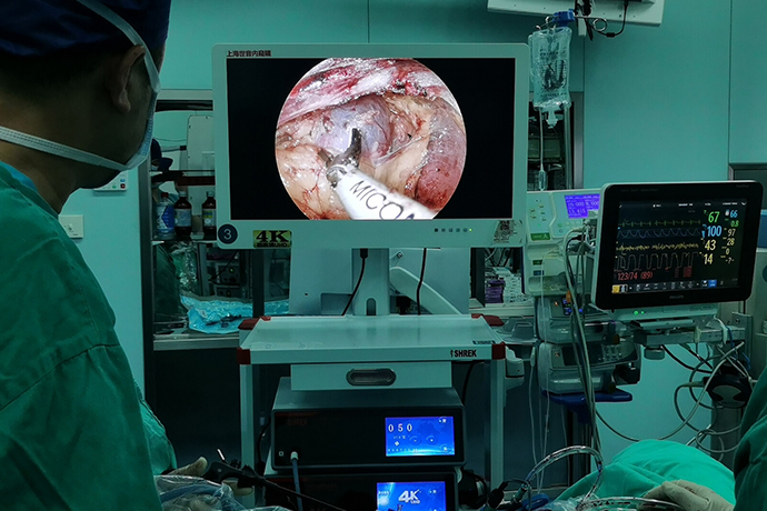
[General Surgery Laparoscopy] 4K ultra-high definition laparoscopic radical nephrectomy
Release time: 18 Jun 2024 Author:Shrek
Kidney cancer, do you know?
Renal cancer, also known as renal cell carcinoma and renal adenocarcinoma, is a malignant tumor originating from the renal tubular epithelium, accounting for 80% to 90% of renal malignant tumors. The incidence of kidney cancer is second only to prostate cancer and bladder cancer, and ranks third among urinary system tumors.
What are the dangers of kidney cancer?
1. Hematuria
It is intermittent painless hematuria, and its severity has nothing to do with the size and stage of the tumor.
2. Pain
About half of the patients have waist pain, mostly dull pain.
3.Block
10% to 30% of patients may develop a mass in the waist or upper abdomen.
4. Extrarenal symptoms
Such as fever, weight loss, anemia, etc., among which fever is the most common.
As surgical procedures become more and more minimally invasive, laparoscopic technology is currently widely used in urology, from the adrenal glands and kidneys to the bladder and prostate. Since the kidneys are located in the retroperitoneum, they are more suitable for posterior 4K ultra-high definition laparoscopic surgery.

4K ultra-high definition laparoscopy technology is an artificially created cavity. Retrolaparoscopic radical nephrectomy has replaced traditional open surgery and become the gold standard for the treatment of localized renal cancer. Retrolaparoscopic radical nephrectomy has the advantages of clear surgical field of view, direct vision operation, precise anatomical level, small trauma, less bleeding, quick recovery, and few complications.
Choice of body position
During retrolaparoscopic radical nephrectomy, the unaffected side folding knife position is used. The operator and assistant are located on the dorsal side of the patient, with the upper limbs abducted and in the natural position. The lower limbs are lowered, and the upper leg is straight and the lower leg is bent.
Things to note include:
1. The patient’s body should be as close as possible to the surgeon, so that the operation will not feel particularly tiring.
2. The monitor is located diagonally opposite the head end, allowing the surgeon and assistant to observe the surgical field at the same time.
3. When the patient is in the folding knife position, there is no need to place a high cushion on the waist. Pay attention to the head and lower limbs not being too low and making an angle of 30 degrees to the horizontal line, which means there is no need to hyperextend the waist. This is different from open surgery. The presence of pneumoperitoneum can better expose the surgical field.
Treatment of renal pedicle blood vessels
The treatment of the renal pedicle blood vessels is the most critical step in retrolaparoscopic radical nephrectomy, and it is also a high-risk operation. There are reports of renal pedicle detachment causing massive bleeding.
The treatment of renal pedicle blood vessels in retrolaparoscopic radical nephrectomy is completely different from that in open surgery. Both operations are performed within the visual range and are firm and reliable. In open surgery, the operation is mostly done by touch, and the treatment of the renal pedicle blood vessels is almost invisible. In retrolaparoscopic treatment of the renal pedicle blood vessels, the arteries and veins are treated separately, while in open surgery, they are treated in a cluster.
When exposing the renal pedicle blood vessels, it is best to dissociate deep between the posterior layer of Gerota's fascia of the kidney and the psoas major muscle to avoid entering the fat sac, maintain the integrity of Gerota's fascia, and facilitate the exposure of the surgical field.
Tips for finding the renal arteries:
1. You can first determine the position of the arcuate ligament, because generally the arcuate ligament points to the location of the renal artery.
2. After finding the reproductive blood vessels, move toward the renal hilum to find the location of the left renal vein. The location of the renal artery is where there are many dense fibers nearby.
3. Observe the pulsation of the artery, and use a suction device to dissociate the area where the pulsation is obvious.
4. If the exposure is not ideal, the patient can be tilted ventrally to increase the exposure field.
When dealing with the renal artery, if it is long enough, it is best to place three hem-lock clips at the proximal end, which is safer.
Placement tips:
1. Place the proximal and farthest ones first, then one or two in the middle, and you'll be fine. And the hem-lock clip must be placed reliably, leaving room for cutting.
2. If the exposure is not ideal, auxiliary holes can be added to facilitate operation.
3. If there is arteriosclerosis, a titanium clip can be added to the proximal end for protection.
The treatment of the renal vein is more difficult than that of the renal artery. Difficulty: The right renal vein is short, the left renal vein has a venous complex, and the vein wall is thin.
Surgical techniques:
1. When treating the right side, it is best to completely free the lower pole of the kidney and slightly elevate it to make the right renal vein longer and easier to handle.
2. When dealing with the right vein, the upper and lower corners of the renal vein must be fully exposed.
3. If the vein is too wide, you can use the edge of a glove or a vascular band to lift the vein and narrow it to facilitate placement of the hem-lock clip.
kidney dissociation
Kidney mobilization is the final stage of retroperitoneal laparoscopic nephrectomy. All surgical stages are indistinguishable. The more last it is, the more attention it deserves. Avoid unnecessary complications.
Surgical techniques:
1. The sequence of dissociation: ventral, lower pole, upper pole, because the dorsal side has been dissociated when dealing with the renal pedicle.
2. Free it outside Gerota’s fascia to ensure the integrity of the fat sac.
3. It is easier to maintain a certain tension when freeing.
4. Maintain the integrity of the peritoneum. If the peritoneum ruptures, do not repair it blindly. If necessary, continue the operation through the peritoneal route.
5. Free the lower pole of the mouse tail first. The advantage is that the upper pole of the kidney has a traction effect. Otherwise, the kidney will fall due to gravity and affect the surgical operation. Finally deal with the upper pole.
6. During the process of freeing the kidneys, the field of view must be within the visual range. The ultrasonic scalpel should be "running at a fast pace" and should not be "big and drastic", and abandon the style of open surgery. Because a little carelessness can have serious consequences. Especially on the right side, there are many injuries to the inferior vena cava.
Adrenal Gland Management
It was previously believed that the scope of radical nephrectomy should include the affected kidney, perinephric fat, perinephric fascia, and ipsilateral adrenal gland. However, current major research results show that routine nephrectomy does not require ipsilateral adrenalectomy.
However, simultaneous ipsilateral adrenalectomy is recommended in the following situations: adrenal gland abnormalities are found in preoperative CT and other imaging examinations or if ipsilateral adrenal gland abnormalities are found during surgery, adrenal metastasis or direct invasion is considered.
Treatment of lymph nodes
Because there is no clear evidence that regional or extensive lymph node dissection in patients with renal cell carcinoma improves overall survival, regional or extended lymph node dissection is not recommended for patients with localized renal cancer.
If obviously enlarged lymph nodes can be palpated during the operation or enlarged lymph nodes are found in preoperative CT and other imaging examinations, enlarged lymph node dissection may be performed to clarify the pathological staging.
Surgical steps
1. Observe the abdominal cavity
2. Open the mesocolon edge and downturn the colon
3. Turn the duodenum inward and downward
4. Free the plane between liver and kidney
5. Open the inferior vena cava sheath
6. Expose the renal veins
7. Free and cut off the genital vein
8. Dissociate the dorsal renal plane upward from the cone-shaped fat at the lower pole of the kidney.
9. Pull the lower pole of the kidney and separate it along the lumbar and dorsal fascia
10. Establish a periureteral plane and cut the ureter
11. Clean lymphatic fat tissue around the renal hilum from bottom to top
12. Free the dorsal plane of the kidney from outside to inside
13. Free and cut the lumbar perforator
14. Free and cut the renal artery
15. Free and cut off the renal vein
16. Establish a plane between the upper pole of the kidney and the adrenal gland
17. Complete removal of the kidney and surrounding tissue
Technical advantages of 4K ultra-high definition laparoscopic radical nephrectomy
1.Postoperative recovery is quick and hospital stay is short.
2.You can eat semi-liquid food the day after the operation, and you can resume your normal life and work one week later.
3.High safety and high quality of life.
4.The surgical trauma is small and the postoperative pain is mild.
5.Generally, patients do not need analgesic treatment after surgery, and the side effects are small.
6.No obvious scars are left and the area is beautiful. Traditional surgical scars are longer, while laparoscopic surgical incisions are more concealed.
7.Accurate and fine, less bleeding. The 4K ultra-high-definition laparoscopic camera has a magnifying effect and can clearly display the fine structure of tissues in the body. Compared with traditional laparotomy, the field of view is clearer, so the surgery is more accurate and precise, effectively avoiding unnecessary damage to organs outside the surgical site. Interference, less intraoperative bleeding, and safer surgery.

- Recommended news
- 【General Surgery Laparoscopy】Cholecystectomy
- Surgery Steps of Hysteroscopy for Intrauterine Adhesion
- 【4K Basics】4K Ultra HD Endoscope Camera System
- 【General Surgery Laparoscopy】"Two-step stratified method" operation flow of left lateral hepatic lobectomy
- 【General Surgery Laparoscopy】Left Hepatectomy