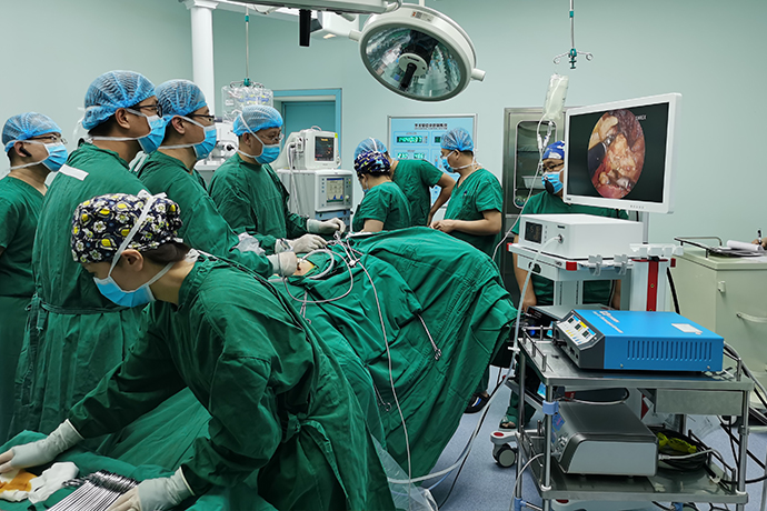
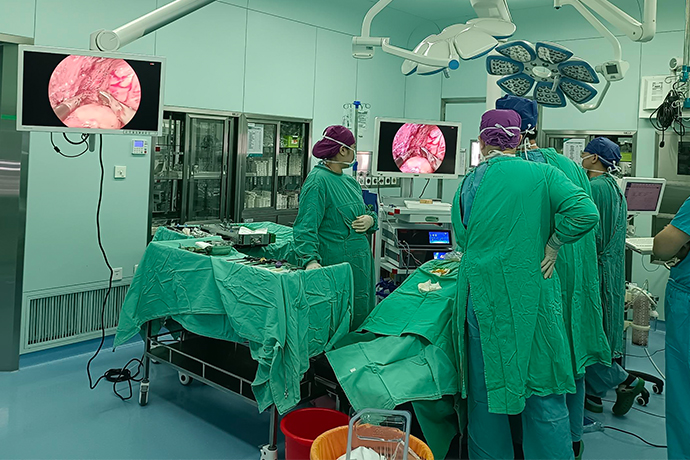
[General Surgery Laparoscopy] 4K ultra-high definition laparoscopic adrenalectomy
Release time: 25 Jun 2024 Author:Shrek
So far, the history of surgical treatment of adrenal glands has been more than a hundred years, and its surgical methods and routes have also undergone continuous development and improvement. In 1992, Ganger et al reported the world's first laparoscopic adrenalectomy (LA), which opened a new chapter in laparoscopic adrenal surgery. Laparoscopic adrenal surgery can significantly reduce intraoperative blood loss, reduce postoperative pain, and shorten hospitalization days, and has greater advantages than open surgery. Laparoscopic adrenalectomy has become the standard surgical procedure for most benign adrenal tumors. In recent years, the development and application of robot-assisted laparoscopy technology in urology surgery has become a general trend. As early as 1999, some scholars performed the first robot-assisted laparoscopic adrenalectomy (RA), and then gradually developed into an important alternative to traditional laparoscopic adrenalectomy. However, the current limited evidence-based medical evidence shows that compared with traditional laparoscopic adrenalectomy, there is no significant difference in perioperative complications, mortality, blood loss, conversion rate, etc. between robot-assisted laparoscopic adrenalectomy and laparoscopic adrenalectomy. But robotic surgery is relatively expensive. Therefore, laparoscopic adrenal surgery will remain one of the important procedures in adrenal surgery.

1. Indications for surgery
Patients with adrenal tumors should consider various factors such as tumor endocrine function, tumor size, tumor nature, and the patient's general condition to determine their surgical approach. Laparoscopic surgery may be considered for benign adrenal tumors less than 6 cm in diameter and some malignant adrenal tumors that are smaller in size. Open surgery is currently the standard surgery for adrenocortical cancer. If the tumor is small and has no invasion of surrounding organs, laparoscopic surgery can also be selected. Most pheochromocytoma can be treated laparoscopically, but in order to avoid local recurrence or seeding caused by intraoperative tumor rupture as much as possible, open surgery is recommended for pheochromocytoma or paraganglioma with a diameter >6 cm or that is invasive; If the surgeon's laparoscopic skills are mature, laparoscopic surgery can also be selected for pheochromocytoma or paraganglioma that is larger than 6 cm and has no obvious invasion of surrounding organs.
For bilateral benign familial pheochromocytoma with MEN2 syndrome, VHL syndrome, NF-1 syndrome, etc., adrenalectomy with partial adrenal tissue preservation should be recommended on the premise of ensuring complete tumor resection. If primary aldosteronism caused by unilateral adrenal hyperplasia or aldosteronoma is diagnosed, unilateral adrenal surgery should be selected, but the specific resection method (total adrenalectomy or adrenalectomy with partial adrenal tissue preservation) is not yet known in the academic community. There is no unified conclusion. For Cushing's syndrome with clear unilateral hyperplasia or adrenal adenoma, it is recommended to consider preserving part of the adrenal tissue on the affected side. For non-functioning adrenal lesions, some normal adrenal tissue should be preserved if the potential for malignancy is not considered.
2. Selection of surgical approach
Adrenal gland (tumor) resection can be done either through the retroperitoneal or transperitoneal approach. Currently, there are different options and advantages for Trocar puncture methods and locations. Here, only one of the methods is selected and introduced.
Retroperitoneal approach
The patient is completely recumbent on the healthy side, in the folding knife position or with a raised waist bridge; a transverse incision is made at two transverse fingers above the iliac crest in the mid-axillary line, and the skin and subcutaneous fat are incised in sequence, and the muscle layer and lumbar tendons are bluntly separated with vascular forceps. membrane, separate the retroperitoneal cavity with fingers, and push the peritoneum ventrally; put the expansion balloon into the retroperitoneal cavity, inflate 600~800 mL, maintain the inflated state for 3~5 minutes, then deflate and pull it out; Two more instrument holes are drilled under the twelfth costal margin in the lower and posterior axillary lines, and 12 mm and 5 mm Trocars are inserted respectively (selected and adjusted according to the surgeon's habits); a 12 mm Trocar is inserted into the original incision as a lens hole, and can be Choose to partially suture the incision to avoid air leakage.
Transperitoneal approach
The patient is placed in a completely normal lateral decubitus position, or in a lateral decubitus position at 60 to 70°, and is placed in a folding knife position or with a raised waist bridge. A pneumoperitoneum is established next to the rectus abdominis at the level of the umbilicus using a Veress needle or small incision, and a 12 mm After Trocar, a 12 mm (left lateral decubitus position) or 5 mm (right lateral decubitus position) Trocar is placed between two transverse fingers below the costal margin at the midclavicular line under direct vision with a laparoscopic monitor, and another trocar is placed near the level of the umbilicus at the level of the front axillary line. The surgeon is accustomed to inserting a 5 mm (left decubitus) or 12mm (right decubitus) Trocar; when in the left decubitus position, a 5 mm Trocar can be inserted under the xiphoid process to lift and stretch the liver during the operation; each Trocar They should be distributed in a triangular shape as much as possible and maintain sufficient distance.
3. Surgical methods and procedures
Insert trocar:
There are many different options for trocar placement, focusing on the three-point puncture method. The first point (camera observation channel) is located at the lateral edge of the right rectus abdominis muscle and at the level of the umbilicus; the second and third points (operating channels) are respectively located on the right anterior axillary line and at the level of the umbilicus, at the lateral edge of the right rectus abdominis muscle and at the level of the umbilicus. 2cm below the costal margin. After first inserting the Veress needle through the first point to establish pneumoperitoneum, insert a 10~12mm cannula from the first point, insert a laparoscopic camera, and explore for organ and intestinal damage, under direct vision guidance Make second and third punctures. The second and third trocars are used for the operating channel. The dominant hand operating channel uses 10~12mm casing; the non-dominant hand operating channel uses 5mm casing. For right adrenal surgery, a fourth trocar can also be inserted to insert straight grasping forceps to push the liver apart. The fourth point can be set at the mid-axillary line and 2cm below the costal margin, and can be adjusted appropriately according to the patient's body shape, tumor size and location.
Expose the adrenal glands and central adrenal vein:
First, incise the lateral peritoneum along the paraascending colic groove, free the ascending colon medially, and expose the perinephric fascia on the surface of the right kidney; then, incise the triangular ligament of the liver, and with the assistance of an assistant, lift the right lobe of the liver upward Push it open so that the entire right lobe of the liver is turned upward to expose the liver surface; then, push the descending part of the duodenum inward to expose the inferior vena cava; finally, open the perinephric fascia and fat sac to expose the right renal hilum. , by dissociating upward along the inferior vena cava, you can see the central vein of the adrenal gland, and from there, you can find the golden adrenal gland in the fat sac of the upper pole of the kidney.
Removal of the adrenal glands (total adrenalectomy):
The inferior vena cava end and the adrenal gland end of the central adrenal vein are clamped and severed with Hem-o-lok, and then the medial edge of the adrenal gland is freed. After freeing the medial edge, the perirenal fat covering the surface of the adrenal gland is lifted up and cut. The perinephric fascia and fat between the adrenal gland and the upper pole of the right kidney are opened. The lateral edge of the adrenal gland is basically avascular, and the entire adrenal gland can be removed after dissociation.
Removal of the tumor (partial adrenalectomy):
If the adrenal tumor is located in the medial branch, lateral branch of the adrenal gland, or the apex of the adrenal gland, partial resection of the adenoma and adrenal gland can be performed. After finding the tumor, use an ultrasonic scalpel to separate the upper and lower edges of the tumor and the front and rear surfaces of the tumor. The adrenal gland tissue connected to the tumor can be cut off with an ultrasonic scalpel or titanium clamps can be used to clip and cut it.
To remove the adrenal gland or tumor:
Carefully explore the surgical field, completely stop bleeding, and place a drainage tube on the wound. Place the removed adrenal gland or tumor into a specimen bag and remove it. The surgical incision is closed layer by layer and the operation is completed.
Left adrenal surgery
Trocar placement: Left adrenal gland surgery generally uses a three-point puncture method. The first point (observation channel) is at the lateral edge of the left rectus abdominis and at the level of the level of the umbilicus. The second and third points are respectively located above the left anterior axillary line and at the level of the level of the umbilicus and 2cm below the lateral edge of the left rectus abdominis and the costal margin. After inserting the Veress needle through the first point to establish pneumoperitoneum, insert a 10~12mm trocar from the first point and insert the laparoscopic camera. After exploring whether there is organ damage in the abdominal cavity, the second and third points of puncture are performed under direct vision guidance. A 10~12mm trocar is placed in the main operating hole; a 5mm trocar is placed in the secondary operating hole. According to intraoperative needs, a fourth puncture point can be made at the posterior axillary line and 2cm below the costal margin, and a 5mm trocar can be inserted to push the kidney or spleen away to expose the location of the adrenal gland.
Expose the adrenal gland and central adrenal vein: First, incise the lateral peritoneum along the lateral parasacral groove of the descending colon, and free the descending colon inward; then, continue to incise upward the peritoneum lateral and above the spleen, and use gravity to move the spleen and pancreatic tail inward. Turn over to expose the perinephric fascia on the anterior medial side of the upper pole of the left kidney; then, cut the renal fascia and fat sac on the medial side of the upper pole of the kidney to find the golden left adrenal gland; finally, expose the left renal pedicle downward. Locate the left renal vein and follow the superior portion of the renal vein to find the central adrenal vein.
Removal of the adrenal gland: The central adrenal vein is freed between the lower edge of the left adrenal gland and the left renal vein, and its renal vein and adrenal gland ends are clamped with Hem-o-lok and then cut. Starting from the upper edge of the adrenal gland, the small branches from the inferior phrenic artery are treated with ultrasonic scalpel or titanium clips; the medial edge is also freed to deal with the small branches from the aorta. There may be some other small blood vessels from the left renal artery and vein at the lower edge of the left adrenal gland, which can be treated with ultrasonic scalpel to reduce bleeding. Finally, the lateral edge is freed and the entire adrenal gland is removed.
Right adrenal gland surgery
1. Establish the level between the upper pole of the right adrenal gland and the liver to reach the uppermost edge of the adrenal gland (this is also the process of making the upper edge of the central adrenal vein).
2. Establish the level of the adrenal gland, the upper pole of the kidney and the renal hilar blood vessels and deepen the level to the muscles (this is also the process of isolating the inferior adrenal artery).
3. Expand the muscle layer cephalad and cut off the middle adrenal artery (this is also the process of establishing the posterior layer of the adrenal ridge and central vein).
4. Open the vena cava sheath between the medial edge of the adrenal gland and the vena cava to expose the front level of the central vein.
5. Disconnect the central vein.
The conventional procedure for organ removal is to cut off the arteries first and then the veins. Reasonable design of surgical steps is that the operation of the previous step pave the way or prepare for the next step or the next few steps. The process of layering is also a process of severing the adrenal artery and exposing the central vein. What finally appears is the ridge where the center of the adrenal gland is located.

- Recommended news
- 【General Surgery Laparoscopy】Cholecystectomy
- Surgery Steps of Hysteroscopy for Intrauterine Adhesion
- 【4K Basics】4K Ultra HD Endoscope Camera System
- 【General Surgery Laparoscopy】"Two-step stratified method" operation flow of left lateral hepatic lobectomy
- 【General Surgery Laparoscopy】Left Hepatectomy