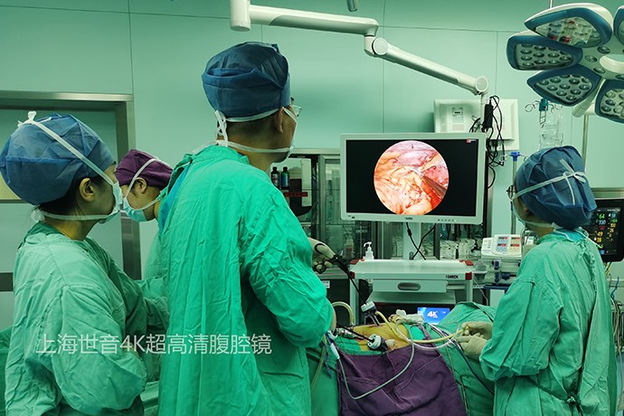
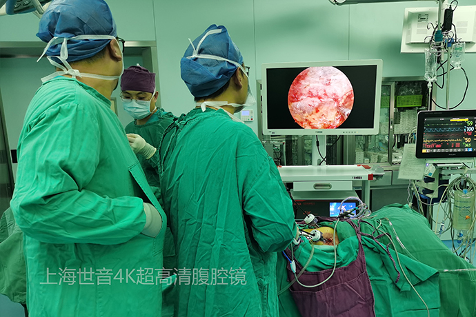
[General Surgery Laparoscopy] 4K ultra-high definition laparoscopic left radical nephrectomy
Release time: 06 Aug 2024 Author:Shrek
Laparoscopic left renal tumor radical surgery is a common surgical method for the clinical treatment of left renal tumors. 4K ultra-high-definition laparoscopy is a minimally invasive surgery with the advantages of less trauma and faster recovery. However, 4K ultra-high-definition laparoscopy for radical resection of left kidney tumors needs to be performed under general anesthesia, which mainly includes the removal of the left kidney tumor and surrounding areas. Lymph node dissection, vascular anastomosis, prosthesis implantation, etc.

The perforator layout for laparoscopic radical nephrectomy via the transperitoneal approach is as follows. Use a small sharp knife to make a small incision at the umbilicus, allowing only the puncture needle to be inserted to establish pneumoperitoneum. Insert the lens puncture device next to the left rectus abdominis and two transverse fingers below the umbilicus. The surgeon's left hand puncture was placed 8cm lateral to the head, and the surgeon's right hand puncture was placed lateral to the foot. The three trocars form an obtuse triangle (green triangle in the picture). Laparoscopic surgery via the transperitoneal approach is slightly different from robotic surgery via the transperitoneal approach. The obtuse angle of the triangle in laparoscopic surgery is smaller, while the obtuse angle of the triangle in robotic surgery is larger (closer to a straight angle).
Choose to insert auxiliary puncture device 1 on the left side of the patient. This puncture device forms an isosceles triangle with the operator's left-hand puncture device and the lens puncture device introduced previously. The layout of the red triangle is crucial for the mobilization of the cephalad part of the kidney during the entire operation. In the dissection of the cephalic part of the kidney, the laparoscopic optical guide rod is inserted through the lens puncture device, and the instruments in the surgeon's left hand (such as bipolar electrocoagulation, aspirators, etc.) For example, ultrasonic scalpel) is inserted through the auxiliary puncture device 1. In the dissection of the foot-side part of the kidney, the laparoscopic optical guide rod is inserted through the lens puncture device, and the instruments in the surgeon's left hand (such as bipolar electrocoagulation, aspirators, etc.) are inserted through the puncture device in the left hand, and the instruments in the surgeon's right hand ( For example, ultrasonic scalpel) is inserted through the puncture device in the surgeon's right hand. Note: During subsequent surgical optimization, the surgeon's right hand puncture should be changed from a 5mm puncture to a 12mm puncture. In addition, the purple auxiliary puncture device can be inserted according to actual needs to determine whether it is necessary.
How do the surgeon and the first assistant (first assistant) choose their standing positions, and how do they allocate the puncture device? The surgeon and an assistant's arms are crossed, and the surgeon's hands insert instruments from the left and right hand punctures respectively. The assistant holds the suction device in his right hand and inserts it through the auxiliary puncture device 1 to assist in exposing and sucking out the oozing blood; the assistant holds the laparoscopic optical guide rod in his left hand and inserts it through the lens puncture device. That is, the first assistant serves as the mirror supporter and the exposure hand at the same time. Because the standing space is limited, it is difficult for the second assistant (second assistant) to assist in the operation.
General steps of surgery:
It is the field of view of the laparoscopic lens entering the abdominal space. Because the tumor is huge, it can be observed under direct vision that the bulging tumor pushes up the left colon and its mesentery.
Find the left paracolic groove and incise the paracolic groove. The purpose is to dissociate the left colon medially and downward to expose the left kidney and left renal tumor that are blocked by it.
After the paracolic groove is incised, the pararenal fat is incised, and the layer between the pararenal fat and the prerenal fascia is freed.
The difficulty in freeing the kidney in this patient is the medial position of the middle and upper pole. There is an adhesion and edema zone between the prerenal fascia ventral to the upper pole of the left kidney and the mesocolon (and the thin pararenal fat below), which is a sign of malignant tumor. High degree of performance. Combined with the surgical video, it can be seen that the adhesions here are serious, the proportion of sharp cuts is higher, and bleeding is easy to occur. In severe areas of adhesion, there are mesocolon tears. At this time, attention should be paid to continuing the correct level of dissection. Return to the level between the prerenal fascia and the mesocolon rather than continuing at the level of the mesocolon breach. Teacher Liu Lei said that difficult and complex surgeries require more basic surgical skills. The specific operations are "exposure", "dissociation" and "stop bleeding". For severe adhesions and edema here, treat them according to the above principles.
On the medial side of the tumor, dissociate along the direction of the left gonad vein from the foot to the head to find and expose the left renal vein. The left renal vein was lifted cephalad, and the main trunk and branches of the left renal artery were freely exposed. Due to the limited operating space, only a single vascular clamp was used to block the two left renal arteries. Strategically, after the kidney is freed, the left renal vein is cut off, and the space is expanded to meet the conditions, the renal artery is blocked and cut off with triple vascular clamps.
The "spleen-standing method" can effectively reduce the difficulty of exposing the head side of the kidney.
The so-called "standing up the spleen", as the name suggests, is to make the spleen "stand up". This is analogous to our previous article introducing the “lifting the roof and shoveling the ground” method for adrenal adenoma. In the past, the difficulty in mobilizing the cephalad side of the left kidney was mainly due to insufficient mobilization of the spleen. As a result, the spleen and pancreas significantly compress the upper pole of the left kidney, resulting in a narrow space.
In order to solve the problem of small space, it requires laborious exposure of the operator's left hand or the assistant's operating lever.
The "spleen-standing method" allows the spleen to "stand up" (just like the "shoveling the ground" method for adrenal adenoma). The spleen will be subject to natural pulling force and hang headward and inward. The space cephalad to the left kidney was naturally exposed. Readers can learn this simple method by watching the surgical video of this stage.
The first step of the "Spleen Standing Method" is to free the splenorenal ligament and connection between the spleen and the kidneys. The first step is to free the width.
The second step of the "Spleen Lifting Method": Below the spleen is the tail of the pancreas, which is dissociated at the level between the pancreas and the kidney. The second step is deep dissociation.
What are the next surgical steps?
1. As the upper pole and medial side of the kidney are freed in depth, the left renal vein is fully exposed. Use vascular clamps to triple clamp and then cut off. After the renal vein was cut, the two renal arteries behind it (which had been blocked by a single vascular clamp) were exposed, and were clamped and cut with triple vascular clamps.
2. It is easy to dissociate the lower pole and dorsal side of the kidney. As shown in the figure below, cut the lateral vertebral fascia on the dorsal side, enter the most familiar level of retroperitoneal surgery, and free the surface along the level of the psoas major muscle. Loose connective tissue. In this case, the dorsal side of the kidney was not invaded by the tumor and the normal renal structure was preserved, so the operation was easy.
3. Because this patient has locally advanced renal cancer and has a high recurrence rate after surgery, continuous titanium clips were used to mark the renal hilum. If the patient requires auxiliary radiotherapy in the future, the titanium clip metal markers will help position the radiotherapy.
4. Connect the incision of the surgeon's left hand puncture tool and the lens puncture tool to serve as the specimen removal port. The incision of the puncture device on the operator's right hand was selected as the drainage tube opening.

- Recommended news
- 【General Surgery Laparoscopy】Cholecystectomy
- Surgery Steps of Hysteroscopy for Intrauterine Adhesion
- 【4K Basics】4K Ultra HD Endoscope Camera System
- 【General Surgery Laparoscopy】"Two-step stratified method" operation flow of left lateral hepatic lobectomy
- 【General Surgery Laparoscopy】Left Hepatectomy