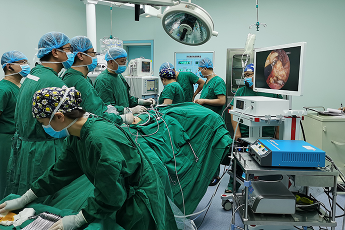
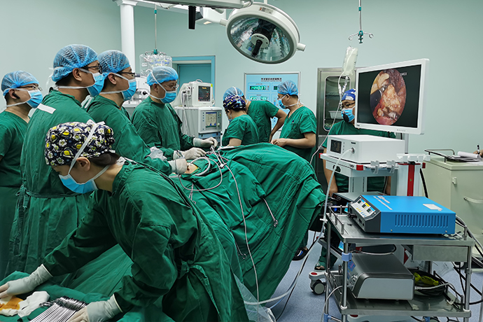
[Orthopedic Arthroscopy] Removal of intra-articular loose bodies
Release time: 15 Oct 2024 Author:Shrek
What is a knee loose body?
Is it necessary to take it out?
Intra-articular loose bodies in the knee joint, also known as "joint mice", refer to movable cartilage or osteochondral fragments in the joint, which is a common knee arthropathy. Intra-articular loose bodies can originate from cartilage, osteochondral or synovial membrane, and can be completely free or connected by soft tissue bands. Clinically, loose bodies in the knee often cause joint locking. Because smaller loose bodies are squeezed between the joint surfaces, sudden joint locking occurs. When it occurs, the patient will have severe pain, and the interlocking position is often not fixed (sometimes in a flexed position, unable to extend; sometimes in an extended position, unable to flex). Patients may experience joint swelling and effusion due to mechanical stimulation of the synovial membrane of the knee joint, making the knee weak or weak, or mobile masses may be touched due to loose bodies traveling to the superficial part. In addition, when locking and unlocking, patients can hear or feel noises and erratic movements, and some may even cause kneeling and falling.

Patients with loose bodies in the knee joint often feel that there are hard objects running around in their knee joints. When walking, sometimes you may suddenly feel that your knee joint is stuck, and you need to move your knee joint before you can extend and bend your knee joint normally.
Loose bodies in the knee joint will wear away the cartilage of the knee joint and gradually expose the subchondral bone, leading to knee osteoarthritis and increasing knee joint pain. Gradually, the knee joint will become deformed, unable to straighten or bend, causing unbearable pain, resulting in the loss of the ability to work and even the ability to walk.
What is the most common cause of loose bodies?
1. Osteoarthritis
It commonly occurs in middle-aged and elderly patients with joint degeneration, aging, and cartilage or osteochondral peeling. Exfoliated tissue accumulates in the joint and is nourished by synovial fluid, gradually forming loose bodies. Osteophytic fractures caused by osteoarthritis hyperplasia can also form loose bodies, especially osteophyte fractures at the upper pole of the patella.
2. Osteochondritis dissecans
Osteochondritis dissecans commonly occurs in young and middle-aged patients. It is caused by ischemic necrosis and detachment of articular cartilage or subchondral bone under the repeated action of external force or inflammatory reaction, forming loose bodies in the joint, which is called osteochondritis dissecans.
3. Patellar luxation
It often occurs in teenagers. At the moment of patellar dislocation, the medial cartilage of the patella and the cartilage of the lateral femoral condyle impact. The impact causes osteochondral damage. The shedding of osteocartilage will form loose bodies, leading to joint nooses.
4. Synovial chondroma
It should be distinguished from loose bodies. This disease is caused by synovial cartilage metaplasia. It is characterized by the formation of cartilage nodules on the synovium. These cartilage bodies can sometimes reach dozens or even hundreds. They can grow with pedicles, protrude into the joint cavity, or fall off into the joint cavity and become loose bodies ( joint rat). Generally, there are <3 loose bodies and >3 synovial chondromas.
Therefore, joint loose bodies can occur in all ages and require detailed analysis of specific medical records. Of course, loose bodies are most likely to occur in middle-aged and elderly patients, with joint degeneration and aging as the risk factors. The knee joint wears and wears, the cartilage softens and falls off, and the subchondral bone is gradually exposed. As a result, knee osteoarthritis occurs, and knee joint pain becomes increasingly severe. Gradually, it will lead to knee joint deformation, pain, and loss of the ability to work and even walk. Therefore, if a loose body is found in the knee joint, it should be removed as soon as possible, otherwise the knee joint will wear out faster and the symptoms and pain will become more severe.
Where are loose bodies commonly found?
Loose bodies most commonly occur in the knees and elbows, and occasionally in the hip joint (femoral head) and ankle joint (talus bone). Among them, because the knee joint cavity is larger and wider, the loose body is easier to obtain. The hip joint is very tight. Without traction, the arthroscope cannot penetrate into the joint cavity at all. What is even more difficult is that loose bodies in the hip joint often appear in the pocket at the inner rear, and the arthroscope and other instruments cannot be bent. Surgery It is necessary to bypass the femoral artery, vein and nerve and can only be inserted from the anterolateral side, so the loose body of the hip joint cannot often be completely removed.
Clinically, loose bodies in the knee often cause joint locking. Because smaller loose bodies are squeezed between the joint surfaces, sudden joint locking occurs. When it occurs, the patient will have severe pain, and the interlocking position is often not fixed (sometimes in a flexed position, unable to extend; sometimes in an extended position, unable to flex). Patients may experience joint swelling, effusion, knee weakness due to mechanical stimulation of the periosteum, or mobile masses may be touched due to loose bodies traveling to the superficial part. In addition, when locking and unlocking, patients can hear or feel noises and erratic movements, and some may even cause kneeling and falling.
Treatment of loose bodies in the knee joint
The treatment of loose bodies in the knee joint can be divided into conservative treatment and surgical treatment.
1. Conservative treatment includes: For small loose bodies that have no obvious noose symptoms and are only found on imaging, conservative treatment can be adopted and oral non-steroidal anti-inflammatory drugs can be used to relieve arthritic reactions and pain. Treatment methods such as elimination of swelling, local hot compress, physical therapy, joint puncture, extraction of fluid accumulation, injection of sodium hyaluronate solution, intra-articular injection of PRP (platelet rich plasma) prepared from autologous blood can relieve and improve symptoms.
2. Surgical treatment: Because minimally invasive arthroscopic treatment has the advantages of less trauma, less pain, less bleeding, faster recovery, and shorter hospitalization time, it has been widely used in clinical practice in recent years.
In this minimally invasive procedure, the surgeon removes fragments from within the knee joint. These fragments are usually a loose piece of bone, cartilage, or other tissue floating in the joint.
In preparation for surgery, anesthesia is administered. Clean the knee joint with disinfectant.
Expose joints
The surgeon makes several small openings in the knee joint. The arthroscope and various arthroscopic instruments are inserted through these openings.
Clean joints
The surgeon carefully examines the joint, looking for signs of damage or debris. The patient may have one or more loose bodies floating in the joint. The surgeon uses a grasping tool to remove debris from the knee joint. If the fragments cause damage to any surface in the joint, the surgeon may also be able to make some repairs.
End of surgery and post-operative care
After the surgery, the instruments are removed, the incision is closed and bandaged. The knee joint will gradually heal over the next few weeks.

- Recommended news
- 【General Surgery Laparoscopy】Cholecystectomy
- Surgery Steps of Hysteroscopy for Intrauterine Adhesion
- 【4K Basics】4K Ultra HD Endoscope Camera System
- 【General Surgery Laparoscopy】"Two-step stratified method" operation flow of left lateral hepatic lobectomy
- 【General Surgery Laparoscopy】Left Hepatectomy