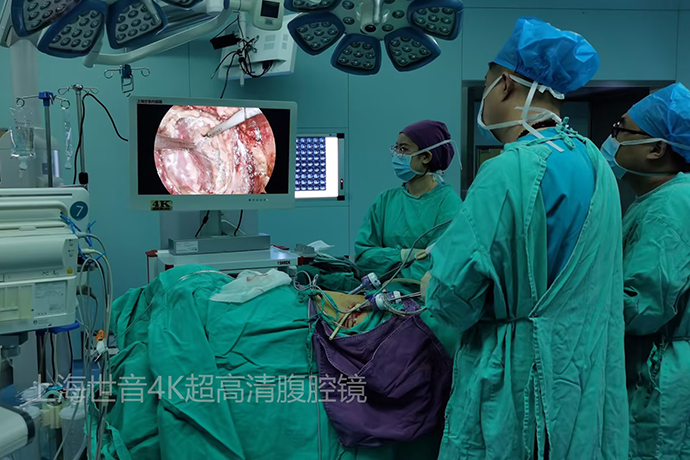
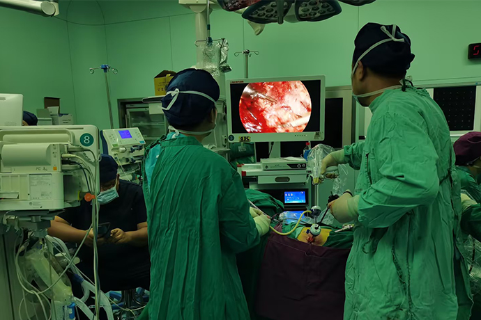
[General Surgery Laparoscopy] Intrahepatic bile duct stones
Release time: 22 Oct 2024 Author:Shrek
Mainly caused by biliary tract infection and cholestasis.
Symptoms such as abdominal pain, high fever, chills, and jaundice are common. Treatment for intrahepatic bile duct stones is mainly surgery.
Early diagnosis and timely treatment are recommended.

Intrahepatic bile duct stones (Hepatolithiasis) are stones located above the Y-shaped bifurcation of the left and right hepatic ducts (i.e., stones in the bile ducts above the confluence of the left and right hepatic ducts). The stones are mostly brown or black in color and are easily Broken, with higher bacterial content. The disease is more common in eastern and southern Asian countries and is a common disease in some areas of my country.
Most frequently asked questions by patients
Are stones stones?
No. Stones do not refer to the stones we see in our lives, but complex calcified deposits formed by the metabolites of various substances in the human body. The main components of intrahepatic bile duct stones are bile pigment stones.
Are intrahepatic bile duct stones easy to remove?
Not easy. Stones are easy to remain after hepatolithiasis surgery, and the incidence rate can reach up to 20% to 40%. In recent years, the rate of residual stones after surgery has dropped significantly. However, some bile duct strictures are difficult to correct at one time and bile duct bleeding during stone removal. The reason is that there is still a certain residual rate of postoperative stones.
Will intrahepatic bile duct stones cause bile duct dilation?
Yes. The liver is a digestive organ. The bile secreted by the liver enters the intestine through the bile duct and participates in the digestion and absorption of food. When the bile duct is blocked by stones, bile cannot be discharged smoothly, and bile continues to accumulate. Increased pressure will cause bile duct dilation.
Will intrahepatic bile duct stones make the liver smaller?
It will. Due to the repeated stimulation of stone obstruction and stagnant bile, biliary cirrhosis will occur, which is manifested as the liver becoming smaller and harder.
Surgical treatment
Minimally invasive surgical treatments for hepatolithiasis include hepatectomy, bile duct lithotomy, and cholangiojejunostomy. The above operations can be completed under traditional laparotomy, laparoscopy or robot-assisted laparoscopy. In principle, the surgical approach is based on diseased liver resection and/or bile duct lithotomy.
liver resection
Under general anesthesia, doctors remove the diseased liver segments, bile ducts and stones together through laparotomy, laparoscopy or the Da Vinci robot. This is currently a relatively effective treatment method.
Indications: Patients with difficult-to-remove stones in diseased liver lobes or bile ducts, difficult-to-correct bile duct stenosis or cystic dilatation, atrophy and fibrosis of liver parenchyma, combined with liver abscess or intrahepatic cholangiocarcinoma.
Bile duct lithotomy
Under general anesthesia, the doctor incises the bile duct to directly remove the stones through laparotomy, laparoscopy or the Da Vinci robot approach, or removes the stones with the help of a choledochoscope.
Indications: There is no obvious atrophy of the diseased liver segment, no severe bile duct stricture, no residual lesions inside and outside the liver after removing all the stones, and multiple stones in the left and right intrahepatic bile ducts. There is a certain recurrence rate after bile duct lithotomy, and some patients may need another operation due to recurrence of stones.
Cholangioplasty and cholangioenterostomy
Under general anesthesia, the bile duct will be anastomosed and connected directly to the intestine through laparotomy, laparoscopy or Da Vinci robotic approach to ensure smooth bile drainage. Indications: Patients with intrahepatic bile duct stones and hilar bile duct stenosis, and the intrahepatic lesions or bile duct stenosis lesions can be removed. In addition, patients with loss of sphincter of Oddi function, cystic dilation of the common bile duct, and cancer of the hepatic hilar and extrahepatic bile ducts may be considered. Use cholangiojejunostomy.
Cholejejunostomy loses the anti-intestinal fluid reflux function of the Oddi sphincter at the lower end of the bile duct, resulting in postoperative complications such as reflux cholangitis, bile duct restenosis, and stone recurrence. Therefore, the indications for surgery should be strictly controlled.
1. Place an indwelling blocking band on the first porta hepatis.
2. Under the controlled low central venous support of the anesthesiology department, the liver was split along the round ligament of the liver to expose the sagittal part of the left Glinsson sheath.
3. Cut off a branch of the S3 segment.
4. The ultrasonic scalpel opens the enlarged and dilated S3 bile duct and removes the stones.
5. The dilated bile duct is closely attached to the left hepatic vein and stripped.
6. Isolate the main trunk of the left hepatic vein and leave a ligature (not ligated yet).
7. Cut off the S2 segment of Glinsson’s pedicle with ultrasonic scalpel.
8. Suspend and ligate the left hepatic vein, and remove the left outer lobe of the liver.
9. Remove the clean stones and close the S2 and S3 bile duct openings with continuous barbed sutures.

- Recommended news
- 【General Surgery Laparoscopy】Cholecystectomy
- Surgery Steps of Hysteroscopy for Intrauterine Adhesion
- 【4K Basics】4K Ultra HD Endoscope Camera System
- 【General Surgery Laparoscopy】"Two-step stratified method" operation flow of left lateral hepatic lobectomy
- 【General Surgery Laparoscopy】Left Hepatectomy