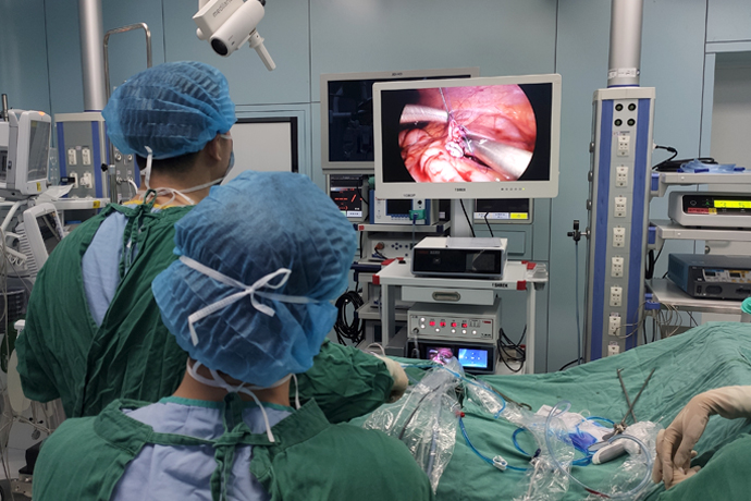
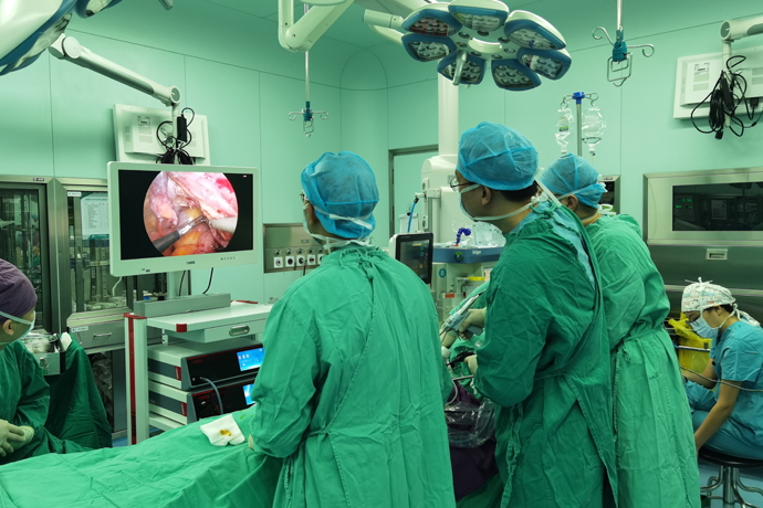
[General Surgery Laparoscopy] 4K Ultra HD Laparoscopic Cholecystolithotomy
Release time: 29 Oct 2024 Author:Shrek
Gallstones are a common disease that is often discovered during physical examinations. There may not be any physical signs at ordinary times, or there may be some related signs, but we are not aware of them. And once gallstones are discovered, is surgery necessary?

When does gallstones require surgery?
With the improvement of people's living standards and changes in dietary structure, the incidence of gallstones has increased than before. Gallstones are further divided into gallbladder stones and bile duct stones. Bile duct stones are further divided into intrahepatic bile duct stones and extrahepatic bile duct stones. Extrahepatic bile duct stones are often combined with gallbladder stones. So what kind of gallstones require surgical treatment?
“Variety” of gallstones
If simple gallbladder stones do not have any symptoms, they are mostly discovered during physical examination and are called "quiet stones". Quiet stones generally do not require treatment.
There is also a view that static stones also need to be treated as soon as possible, because long-term stone stimulation can induce gallbladder cancer.
Surgical treatment is recommended for gallbladder stones with acute and chronic inflammation, larger stones, gallbladder polyps, and complications such as biliary pancreatitis.
According to the guidelines, laparoscopic cholecystectomy is the preferred treatment for gallbladder stones. For those who cannot tolerate surgery, gallbladder puncture and drainage can be considered.
For patients with gallbladder stones combined with common bile duct stones, early treatment is recommended.
There are two main treatment methods: laparoscopic cholecystectomy + ERCP lithotomy and laparoscopic cholecystectomy combined with choledocholithotomy. Both methods have their own advantages and disadvantages, and individualized treatment plans should be formulated according to the specific conditions of the patient.
Specific steps for gallbladder stone surgery
STEP 1: Make the first incision at the umbilicus, insert the cannula, and put in the camera.
STEP 2: Make the remaining three incisions in the upper abdomen and insert the cannula.
STEP 3: Find the gallbladder and grab it with clamps.
STEP 4: Peel off the fat to reveal two important structures - the cystic duct and the cystic artery.
STEP 5: Clamp and cut the cystic duct and cystic artery.
STEP 6: Begin to separate the gallbladder from behind the liver.
STEP 7: Carry out gentle movements until the gallbladder is completely peeled off.
STEP 8: Put the gallbladder into this small bag and take it out from the incision in the upper abdomen.
STEP 9: Gallbladder stones.
Key points and difficulties of laparoscopic cholecystectomy
Good surgical field exposure is the prerequisite for correct treatment of structures within Calot's triangle. Correct treatment of structures within Calot's triangle is the key to complete cholecystectomy. The quality of treatment is directly related to the patient's prognosis.
1. Well exposed Calot triangle
①After inserting a laparoscope at the abdominal wall puncture position to understand the height of the liver, make a puncture hole vertically or slightly below the lower edge of the liver on the right side of the falciform ligament under the xiphoid process. The distance should be about 10cm, and the puncture holes under the costal margin of the midclavicular line and anterior axillary line should be slightly lower than the lower edge of the liver.
②Changes in body position and pneumoperitoneum pressure. For those with a lot of intra-abdominal fat and poor intestinal preparation and flatulence, the upward movement of the greater omentum and gastrointestinal tract will shrink the subhepatic space and poor exposure of the Calot triangle. At this time, appropriate Increase the intra-abdominal pressure to 15mmHg. During the operation, the patient is placed in a head-high and feet-low position and tilted 15 degrees to the left. With the help of the gravity of the above-mentioned organs and fat tissue, the subhepatic space is widened and the operating space is increased.
2. Dissect and process tissues within Calot’s triangle
Calot's triangle should be opened as much as possible, and the surrounding area around the confluence of the ampulla of the gallbladder and the cystic duct should be fully freed to clearly display the structures within it. You may encounter the following problems during surgery:
① If the adhesions in the Calot triangle are severe, the following points should be noted during separation: The adhesions should be separated close to the ampulla of the gallbladder under tension and traction, and the operation should be gentle. Blunt separation methods (such as separation forceps, uncharged electric hooks) are generally used. and micro electric clippers, etc.) to avoid blind electrocoagulation and electrocution. Cases of extrahepatic bile duct injury due to thermal burns have been frequently reported.
In cases of acute cholecystitis (or with stone incarceration), there is often obvious congestion and edema in Calot's triangle, but the adhesion is not serious. There is often extravasation of edema fluid when isolating the ampulla of the gallbladder and dissecting Calot's triangle. You can use the head end of the irrigator to bluntly separate the gallbladder ampulla and dissect Calot's triangle while rinsing repeatedly to keep the field of vision clear, and the operation is more likely to be successful.
If there is no gap adhesion between the common bile duct, common hepatic duct, right hepatic duct and the ampulla of the gallbladder, the serosa can be incised at the junction of the ampulla of the gallbladder and the cystic duct, and separated above the cystic duct with dissecting forceps to display Calot's triangle. If the cystic duct cannot be exposed, retrograde resection of the gallbladder can be used.
②The anatomical variation in thickness and length of the cystic duct is large, and the following principles should be followed when exposing and processing it: the surrounding area around the junction of the cystic duct and the ampulla of the gallbladder must be fully freed. If the "three tubes and one ampulla" (liver Common duct, common bile duct, cystic duct, gallbladder ampulla), when clamping the cystic duct, there should be an empty space above the junction of the cystic duct and the common hepatic duct (meaning that the common hepatic duct is not in it).
After the cystic duct is clamped, it should be cut off to avoid extrahepatic bile duct burns due to heat. If the cystic duct is obviously thickened and direct treatment is difficult, the gallbladder can be resected retrogradely and ligated with a snare.
③There are many anatomical variations in the shape and branches of the cystic artery. It is important not to deal with one branch while neglecting other branches. It is best not to "skeletonize" the cystic artery to avoid insufficient vascular tissue and unstable clamping. When the separated gallbladder bed encounters a larger vascular branch, clamping should also be performed to stop bleeding, especially when there is no main cystic artery in Calot's triangle.
Pay attention to whether there is an artery behind the cystic duct. This situation should be suspected if there is an obvious feeling of toughness behind the cystic duct when separating the cystic duct. At this time, the upper half of the clamped cystic duct can be cut off first, and then the distal part of the cut cystic duct can be repaired. Use a clamp, and then cut off the entire cystic duct (including the cystic artery behind the cystic duct).

- Recommended news
- 【General Surgery Laparoscopy】Cholecystectomy
- Surgery Steps of Hysteroscopy for Intrauterine Adhesion
- 【4K Basics】4K Ultra HD Endoscope Camera System
- 【General Surgery Laparoscopy】"Two-step stratified method" operation flow of left lateral hepatic lobectomy
- 【General Surgery Laparoscopy】Left Hepatectomy