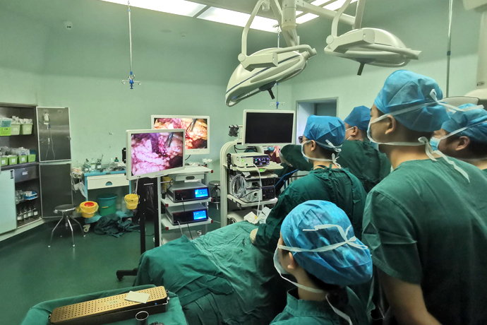
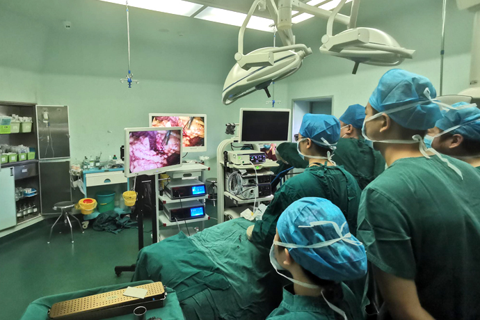
[General Surgery Laparoscopy] 4K Ultra HD Laparoscopic Surgery for Acute Cholecystitis
Release time: 05 Nov 2024 Author:Shrek
Cholecystitis, including acute cholecystitis and chronic cholecystitis, is an inflammatory reaction in the gallbladder caused by gallstones or other causes.

The main cause of this disease is infection caused by biliary obstruction and cholestasis. Biliary stones are the main cause of biliary obstruction. Cholecystitis is a common digestive system disease. The high-risk groups are mainly middle-aged and elderly people, and it is related to gender, eating habits and other factors. The incidence of acute cholecystitis is higher in women.
The main symptoms of cholecystitis include pain in the right upper abdomen, which may be accompanied by nausea, vomiting, anorexia, and constipation. Among them, acute cholecystitis develops rapidly, and serious complications such as gallbladder perforation may occur when the disease worsens. Cholecystitis is not contagious.
1. Introduction
Acute cholecystitis is one of the most common causes of acute abdominal pain. Decisions must be made promptly.
The use of laparoscopy for acute cholecystitis is increasingly less controversial. In fact, laparoscopic cholecystectomy is now increasingly used for the treatment of acute cholecystitis, with advantages including faster recovery and shorter hospital stay compared to open cholecystectomy.
2.Dissection
Anatomy
1. Make the first incision at the umbilicus, insert the cannula, and put in the camera.
2. Make the remaining three incisions in the upper abdomen and insert the cannula.
Regional anatomy
1. Liver
2. Stomach
3. Lesser omentum
4. Gallbladder
5. Hepatic flexure
6. Greater omentum
Regional anatomy
1. Bottom
2.Body
3. Funnel
4.Cholecystic duct
5. Common hepatic duct
6. Common bile duct
Vascular supply
1. Gallbladder
2.Cholecystic artery
3. Mascagni lymph nodes
4. Proper hepatic artery
5. Abdominal aorta
6.Portal vein
7.Gastroduodenal artery
3. Pathological anatomy
In AC, there is acute inflammation of the gallbladder wall due to long-term obstruction of the cystic duct or neck of the gallbladder. It can also occur as an inflammatory response transmitted by stones.
Inflammatory adhesions can often be found around the gallbladder to adjacent organs such as the duodenum, right colon, and greater omentum.
We distinguish:
Catarrhal acute cholecystitis.
Purulent cholecystitis.
Gangrenous cholecystitis.
4. Anatomical variations I
cystic artery changes
The anatomy of the biliary vasculature is highly variable from patient to patient, particularly with the right hepatic and cystic arteries.
A good working knowledge of the various abnormalities that may be encountered will help identify important structures and prevent intraoperative complications.
Double cystic artery
Variation 1
Dual cystic arteries; both arise from the normal right hepatic artery in the gallbladder triangle.
Variation 2
Two cystic arteries; one posterior-inferior and one anterior-superior to the cystic duct
Variation 3
Both cystic arteries and cystic duct are superior to cystic duct.
origin of gallbladder artery
Variation 1
The gallbladder artery originates from the proper hepatic artery
Variation 2
The cystic artery originates from the normal left hepatic artery, the gallbladder triangle
Variation 3
The cystic artery originates from the celiac trunk, anterior superior - cystic duct
5. Anatomical Variations II
intrahepatic bile duct
1. Common bile duct
2. Gallbladder
3.Cholecystic duct
4. Right hepatic duct
5. Left hepatic duct
Right hepatic duct I
repeat
Unique right hepatic duct (53% cases)
Duplicate malformation of right hepatic duct (47% cases)
RL: Right tube
Turn: The tube in the middle right
Branch
Upper bile duct bifurcation (10% cases)
Right paralateral (anterior) duct Right (posterior) left hepatic duct
Tail entrance of RL pipe
The right (rear) duct of the caudal entrance enters the main channel (6% of cases)
Tail inlet of RPM pipe
Right paramedian caudal entry (anterior) catheter into the main channel (20% of cases)
6. Anatomical variations III
Changes in extrahepatic bile ducts
A sound, working knowledge of anatomical variations will facilitate intraoperative identification of various ductal structures. In addition, strict compliance with the basic rules of exposure and dissection, as well as mastery of laparoscopic techniques, will further protect against potentially serious complications of the surgical procedure.
7. Anatomical variation IV
morphological factors
The patient's morphological characteristics may require adaptation of the basic technique.
Hypertrophy of the right lobe of the liver or an overly large gallbladder can present difficulties during dissection. In these cases, the position of the retraction needle can be adjusted to allow improved access to the subhepatic region.
8.Indications
Clinical symptoms of inflammation on admission:
High fever above 37.5℃;
nausea or vomiting;
Severe pain in the right lower or upper abdomen.
Ultrasound evidence:
Thickened gallbladder wall (>=4mm);
dilated gallbladder
American sign of Murphy's sign when ultrasound probe was introduced.
Relative contraindications
Patients with ASA 4 (moribund) or acute calcific cholecystitis in the intensive care unit (percutaneous cholecystotomy may be a good option for these critically ill patients).
Gangrenous cholecystitis (abscess).
Long-term AC (10+ days): This remains controversial as many recent reviews found early laparoscopic cholecystectomy to be safe due to shorter hospital stay, improved quality of life, and improved quality of life compared with delayed laparoscopic cholecystectomy is not accompanied by more complications (Wilson E et al., 2009).
Surgeon's limited experience in laparoscopy.
Operation time:
It has been debated for many years that surgical approaches for the treatment of acute cholecystitis, whether surgical (open or laparoscopic), should be performed early or delayed after the onset of symptoms. Currently, most studies suggest that, on average, early laparoscopic cholecystectomy is less expensive than delayed approaches. Outcomes regarding quality of life appear to improve (Wilson E et al, Br J Surg, 2009). Regarding surgical outcomes, laparoscopic treatment of acute cholecystitis was associated with an increased conversion rate (10–15% vs. 1–2%) but not with a higher incidence of intraoperative or postoperative complications (Michalowski K et al ., Br J Surg, 1998; Csikesz NG et al., Surgery, 2008).
9. Operating room setup
Patients are prepared and covered in the usual way:
Standard skin preparation;
sterile area;
Patient placement:
supine position;
Left arm 90°;
Right arm along body;
Leg abduction.
Team
1. The surgeon is positioned between the patient’s legs.
2. The first assistant is on the left side of the patient.
3. The second assistant is usually on the patient's right hand, opposite the first assistant.
Equipment
1. Radioactive equipment (optional)
2. Laparoscopic group
3. Anesthesia machine
4. Laparoscopic unit (optional)
5. Instrument panel
6. Electric iron
7. Operation desk
10. Trocar puncture
In most cases, the 4-hole method is used. By converting to an open approach, acute cholecystitis may be associated with intestinal obstruction or adhesions.
Optics
The optical port is placed at the umbilical area (or higher in obese patients).
Operate
Two operating cannulas were placed in the right upper quadrant and left subcostal area. Introduce graspers, scissors, hooks, dissectors and clamps through these trocars.
Tractor
The retractor trocar is placed in the upper abdominal area.
Through it the suction and irrigation device is introduced.
Optional
A fifth optional trocar can be placed midway between the umbilical trocar and the left subcostal trocar. It is used to pull on the duodenal structures caudally.
When this fifth optional trocar is added, the upper abdominal trocar is used for aspiration and irrigation devices.
11.Instruments
Optics
In addition to a direct looking lens (0°), it is also necessary to have an observer with a large depth of field combined with an HD camera. The use of a high-quality camera is recommended as it significantly increases the possibility of anatomical identification in difficult conditions (Singhal T et al., 2009).
Operating equipment
Most surgical teams use instruments with a diameter of 5mm.
Take back equipment
Atraumatic grasping forceps must be used. Suction devices are often used as retractors.
Optional equipment
The effectiveness of ultrasonic dissectors on inflamed tissue remains to be demonstrated.
12. Main principles
The purpose of this surgical technique is to remove the gallbladder after freeing it from adhesions to adjacent organs.
Compared with elective cholecystectomy, certain technical points must be emphasized:
1. Decompression of the gallbladder so that it can be optimally grasped.
2. Dissection in contact with the gallbladder wall.
3. Use ultrasonic cautery with caution.
4. Frequent suction and irrigation of the surgical field.
5. Subtotal cholecystectomy remains controversial in difficult cases: however, for others it remains an option in cases of extensive inflammation or fibrosis, which can preclude routine dissection of the Calot triangle (Michalowski K et al., 1998; Singhal T et al., 2009).
6. Systematic use of extraction bags.
7. Drainage of the gallbladder bed (controversy).
13. Exploration
Check for peritoneal effusion
Thorough exploration of the abdominal cavity is required to detect peritoneal fluid and associated lesions.
Abdominal exploration should assess:
Inflammatory state and possible necrosis of the gallbladder,
Inflammation spreads to the hepatoduodenal ligament (porta liver).
It should search for:
The presence of adhesions around the gallbladder (to the omentum, duodenum, colon, etc.),
Collections of bile or pus are present.
14.Exposed
in principle
Exposure of the surgical site is the first step in all surgical procedures. In AC, this procedure is made difficult by inflammatory adhesions to adjacent organs. First, the gallbladder must be in the right subhepatic zone and then at the level of Calot's triangle.
Right subhepatic area exposed
3 techniques
To open the right subhepatic area (difficult operation due to inflammatory adhesions), 3 techniques are used:
suspension of liver;
Using gravity, this naturally lowers the organ toward the pelvis;
Use retractors on adjacent organs.
Suspension
Percutaneous penetrating sutures are used to elevate the liver above the round ligament, improving exposure of the Calot triangle and hepatic pedicle. It passes beneath the ribs and to the right of the abdominal wall of the suspensory ligament. It runs through the lower edge of the round ligament of the liver, and the line pulls out the left side of the suspensory ligament, and its angle is close to that of the suspensory ligament. Tighten the gauze over the gauze pad on the outside of the abdominal wall.
Gravity
The patient is placed in a steep reverse position with a slight left tilt, causing the organs to descend into the pelvis and to the left side.
Retract
Due to the fragile inflammatory state of the tissue, retraction of adjacent organs must be atraumatic.
Gallbladder neck exposed
Principle
After release of adhesions, exposure of the gallbladder neck is possible. This can be achieved after decompression of the gallbladder, which allows the surgeon to properly grasp the gallbladder wall.
Reduced pressure
Using a needle (e.g., Veress needle) inserted laterally under the ribs and through the base of the gallbladder, the gallbladder is emptied to obtain a bacteriological sample (routine culture) and enables appropriate grasping with noninvasive forceps.
Release
Release of adhesions is easily achieved within the first 3 to 4 days of inflammation. Most adhesions involve the omentum, but they can also involve the duodenum and right colon. Ideally, anatomical planes should be found and attempts should be made to minimize unnecessary bleeding.
15. Anatomy
Calot's triangle
Dissecting Calot's triangle is the most delicate step of the operation. Access to the infundibulum is often difficult because stone incarceration displaces it toward the hepatoduodenal ligament. Inflammation can induce adhesions between these structures.
Anatomy progress
1. Funnel
2. Bile duct
Once the perigallbladder inflammatory adhesions have been removed, dissection of Calot's triangle always begins at the suspected junction between the gallbladder neck and the cystic duct. It should not start at the junction of the cystic duct and common bile duct (CBD) because the cystic duct is sometimes very short and can therefore be confused with CBD.
Dissection of the cystic duct is performed 1 cm from the gallbladder neck toward the CBD. When dissecting Calot's triangle, contact with the gallbladder wall is necessary.
Technical insight
Depending on the surgeon's preference, he or she can choose from a variety of instruments for dissection: monopolar hook electrodes, ultrasonic scalpels or hooks, monopolar scissors, dissectors, or peanut-like swabs.
In AC, the presence of inflammatory tissue always makes the surgical area bleed more. Therefore, the following measures need to be taken:
Use a high-performance light-enhanced HD camera: The use of a high-quality camera is recommended as it significantly increases the possibility of anatomical identification in difficult situations.
Frequently irrigate and clean the surgical area with a suction device.
Danger
Electrocautery should only be used selectively and with great caution. In the case of diffuse outflow from inflamed tissue, the conductivity increases due to the water content and the efficiency of electrocautery decreases. The danger therefore lies in increasing the electrical power in contact with the biliary tract (risk of secondary necrosis or stricture).
Mutations
In cases where a large stone is embedded in the gallbladder neck making access to Calot's triangle difficult, after aspiration of the gallbladder contents, the gallbladder neck can be opened and the stone removed and placed in a plastic bag introduced through the abdominal cavity Pass the trocar through the bag.
Once the surgeon has identified the location of the cystic duct from within the gallbladder lumen, dissection of the cystic duct and superior portion of Calot's triangle can begin. This is particularly useful in cases of inflammatory adhesions between the gallbladder and hepatoduodenal ligament.
16.IOC: Technology
Indications
This is indicated when the presence of CBD stones is suspected.
It is used for:
Determine the location, size and number of stones,
Assessing intrahepatic and extrahepatic bile duct anatomy: anatomical variations and CBD size.
Cystic duct incision
Cholangiography was performed through a semicircular incision along its anterior surface to the cystic duct approximately 1 cm from its junction with the CBD. A cystic ductotomy is performed to avoid problems with bile duct insertion due to valves or folds in the cystic duct. The right margin of the CBD must be determined.
The location of the console
The console is brought back to a flat position (i.e., removed from reverse head-down supine position and left tilt) and tilted slightly to the right to displace the CBD anteriorly.
Cholangiography
Catheter introduction
The biliary catheter is brought to the cholecystotomy site percutaneously or via a right subcostal trocar using a rigid introducer. It is inserted 1 to 3 cm into the cystic duct and held in place with a clamp or grasper.
Cholangiography should be performed in three steps:
1. Under radiological guidance, a few milliliters of dilute contrast medium are injected into the bile duct. Static cholangiography can detect CBD stones.
2. Continue dye injection until a complete cholangiogram is obtained. A second radiograph is performed to confirm. The head-down supine position promotes opacification of the intrahepatic bile ducts.
3. The passage of dye into the duodenum at low pressure should be confirmed by a third radiograph.
17. Ligation and segmentation
method
The surgical procedure is delicate and often hemorrhagic due to inflammation of the tissue. Sometimes there is no anatomical plane between the liver and gallbladder. Routinely, cholecystectomy is performed using a retrograde approach, but this will depend on the surgical findings and difficulties, which may require a change in strategy.
18.Extraction
At the end of the procedure, the gallbladder is placed in a protective extraction bag to avoid any contamination of the abdominal wall during extraction.
After increasing the left subcostal trocar incision from 10/11 mm to 2 to 3 cm, extraction was most often performed in the left subchondral area. Due to the 2-layer closure, it is preferred to the umbilical cord incision, which seems to be of better quality and more reliable.
19. End of program
Examine
Before closing the abdominal wall, the surgeon must verify that there is no abnormal bleeding or bile leakage in the gallbladder bed area and that the infrahepatic and infradiaphragmatic spaces have been appropriately lavaged.
lavage
The gallbladder bed is flushed with physiological solutions to remove clots from the surgical area and look for bile leaks or bleeding. The subdiaphragmatic area must also be irrigated to prevent subphrenic abscess.
Drainage
Drainage of the right lower liver region can be performed using a 12 to 15 French multi-perforated silicone drainage tube (with its tip touching the hepatoduodenal ligament) to prevent blood clots in the gallbladder bed and to drain any postoperative bile leakage.
SFCD recommendations (Mutter, 1999)
After cholecystectomy for acute cholecystitis, drainage of the liver bed is not mandatory but should be decided on a case-by-case basis based on the degree of hemostasis and bile stasis.
Closure
After ensuring that no parietal bleeding is present, the trocar can be removed under direct vision and the trocar incision is closed.
For 5mm incisions, only the skin is closed. Incisions measuring 10 mm or larger are closed at both the deep myofascial and skin layers.
20. Postoperative management
If there is no abnormal bleeding or bile leakage, remove the abdominal drainage tube 1 or after surgery.
Early ambulation is necessary (same day of surgery or the day after).
Return to normal eating habits is usually on day 1 after surgery.

- Recommended news
- 【General Surgery Laparoscopy】Cholecystectomy
- Surgery Steps of Hysteroscopy for Intrauterine Adhesion
- 【4K Basics】4K Ultra HD Endoscope Camera System
- 【General Surgery Laparoscopy】"Two-step stratified method" operation flow of left lateral hepatic lobectomy
- 【General Surgery Laparoscopy】Left Hepatectomy