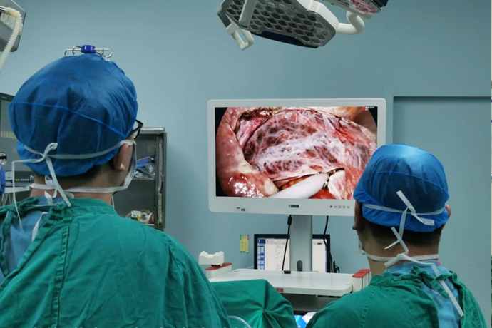
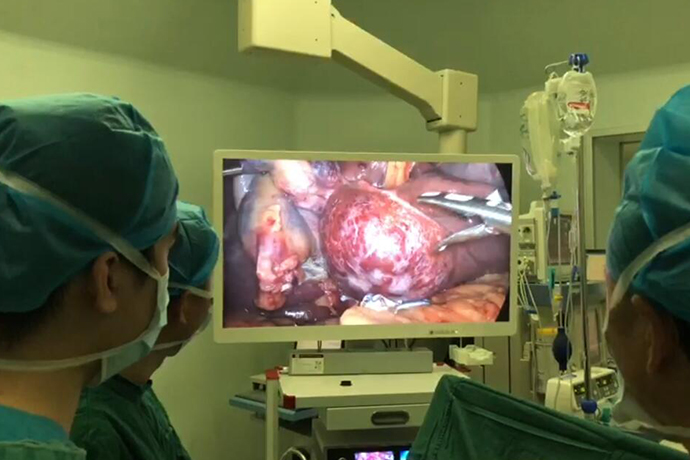
[General Surgery Laparoscopy] 4K ultra-high definition laparoscopic pancreatic body and tail resection treatment
Release time: 12 Nov 2024 Author:Shrek
Indications for body and tail resection surgery
Laparoscopic pancreatectomy is suitable for the treatment of distal pancreatic tumors, including spleen-preserving pancreatectomy and non-spleen-preserving pancreatectomy. Spleen-preserving pancreatic distal resection is mainly suitable for the treatment of benign tumors and low-grade malignant tumors in the pancreatic body and tail; non-spleen-preserving pancreatic distal resection is suitable for radical resection of distal pancreatic cancer and distal pancreatic cancer that cannot preserve the spleen. Treatment of benign tumors.

Main indications for laparoscopic pancreatectomy
Various benign tumors in the body and tail of the pancreas: pancreatic endocrine tumors, serous cystadenoma, etc.
Various borderline tumors in the body and tail of the pancreas: borderline mucinous cystadenoma, intraductal papilloma, etc.
Various malignant tumors in the body and tail of the pancreas, with mild adhesion to surrounding tissues and no distant metastasis according to preoperative assessment, such as pancreatic cancer.
Pancreatic damage, chronic pancreatitis.
Pancreatitis combined with pseudocyst.
Indications for laparoscopic spleen-preserving distal pancreatectomy
Benign tumors and low-grade malignant tumors in the body and tail of the pancreas, such as pancreatic cystadenoma, endocrine tumors, cystadenocarcinoma, etc.
The lesions are mainly concentrated in the left half of the pancreas and the symptoms are chronic pancreatitis.
Chronic pancreatitis combined with pancreatic body and tail cysts.
Indications for laparoscopic distal pancreatectomy without spleen preservation
In summary, it applies to two situations: one is a malignant tumor, and the spleen must be removed in order to ensure the thoroughness of tumor treatment (the spleen must be removed); the other is a lesion that affects the blood supply of the spleen and is urgently removed (the spleen must not be removed). Specific indications are as follows:
Various malignant tumors of the pancreas.
Various borderline tumors of the pancreas, those that are large or have invaded the splenic blood vessels.
The tumor has involved the splenic blood vessels, is close to the splenic hilum, or is adherent to the splenic hilum.
The pancreas has severe chronic inflammation and is tightly adherent to the splenic blood vessels. Forcible separation may lead to fatal bleeding.
The tumor is larger, with a diameter greater than 5.0cm.
Surgical steps
1. Trocar layout: A 5 mm Trocar is inserted into the umbilicus. After entering the scope, under laparoscopic monitoring, the Trocar is inserted under the costal margin of the right anterior axillary line, the lateral edge of the right mid-abdomen rectus abdominis, and the lateral edge of the left upper quadrant rectus abdominis.
2. Exploration and exposure of the pancreas: The gastrocolic ligament was incised with the ultrasonic scalpel, and a large amount of pancreatic juice and bloody fluid accumulated in the lesser omental sac. There was obvious edema in the middle part of the pancreas. The pancreas was completely ruptured due to separation and adhesion, and there was overflow of pancreatic juice and active bleeding in the broken end. Exploration showed that the main trunk of the splenic vein was broken, making repair difficult.
3. Treatment of the splenic vein at the splenic hilum: The splenic hilum is slightly freed at the pancreatic tail, and the main splenic vein before entering the splenic hilum is ligated and severed.
4. Exposure of the splenic artery: Free the tail of the pancreas at the splenic hilum and turn it to the right to protect and free the splenic artery until the broken end of the body of the pancreas. Pay attention to the small branch arteries from the splenic artery to the tail of the body of the pancreas.
5. Cut off the pancreas and process the cross section. Use an ultrasonic scalpel to cut off the body of the pancreas and use bipolar electrocoagulation to stop bleeding. Use 4-0 Prolene sutures to perform interrupted suturing of the proximal pancreatic section. Be careful not to suture the splenic artery.
6. Hemostasis and drainage: After the wound has been completely hemostatic, put the pancreatic tissue into a specimen bag, slightly expand it through the umbilical hole and pull it out. Place a drainage tube near the pancreatic section and lead it out through the left Trocar hole.
Laparoscopic pancreatectomy is suitable for the treatment of distal pancreatic tumors, including spleen-preserving pancreatectomy and non-spleen-preserving pancreatectomy. Spleen-preserving pancreatic distal resection is mainly suitable for the treatment of benign tumors and low-grade malignant tumors in the pancreatic body and tail; non-spleen-preserving pancreatic distal resection is suitable for radical resection of distal pancreatic cancer and distal pancreatic cancer that cannot preserve the spleen. Treatment of benign tumors.
Main indications for laparoscopic pancreatectomy
Various benign tumors of the body of the pancreas: pancreatic endocrine tumors, serous cystadenoma, etc.
Various borderline tumors in the body and tail of the pancreas: borderline mucinous cystadenoma, intraductal papilloma, etc.
Various malignant tumors in the body and tail of the pancreas, with mild adhesion to surrounding tissues and no distant metastasis according to preoperative assessment, such as pancreatic cancer.
Pancreatic damage, chronic pancreatitis.
Pancreatitis combined with pseudocyst.
Indications for laparoscopic spleen-preserving distal pancreatectomy
Benign tumors and low-grade malignant tumors in the body and tail of the pancreas, such as pancreatic cystadenoma, endocrine tumors, cystadenocarcinoma, etc.
The lesions are mainly concentrated in the left half of the pancreas and the symptoms are chronic pancreatitis.
Chronic pancreatitis combined with pancreatic body and tail cysts.
Indications for laparoscopic distal pancreatectomy without spleen preservation
In summary, it applies to two situations: one is a malignant tumor, and the spleen must be removed in order to ensure the thoroughness of tumor treatment (the spleen must be removed); the other is a lesion that affects the blood supply of the spleen and is urgently removed (the spleen must not be removed). Specific indications are as follows:
Various malignant tumors of the pancreas.
Various borderline tumors of the pancreas, those that are large or have invaded the splenic blood vessels.
The tumor has involved the splenic blood vessels, is close to the splenic hilum, or is adherent to the splenic hilum.
The pancreas has severe chronic inflammation and is tightly adherent to the splenic blood vessels. Forcible separation may lead to fatal bleeding.
The tumor is larger, with a diameter greater than 5.0cm.
Pathological diagnosis
Edema can be seen in the pancreatic tissue, vasodilation, congestion and bleeding can be seen around the pancreas, and a small amount of lymphoid tissue can be proliferated.
Postoperative follow-up
B-ultrasound showed (one week after surgery): the portal vein was full of blood. The main trunk of the splenic vein is not shown, but a blue blood flow can be seen twisting along the upper part of the left kidney and shunting a blue blood flow to the left adrenal gland.
The pancreatic cross-section drainage tube was removed on the 3rd day after the operation. B-ultrasound monitoring on the 7th day after the operation showed the formation of splenorenal shunt blood vessels. The patient was cured and discharged on the 11th day after the operation. She has been followed up for 8 months and her growth and development is normal.
Key points of surgery
1. For pancreatic damage caused by trauma, surgical exploration should be performed as soon as possible, otherwise local edema, severe inflammatory reaction, and severe bleeding will affect the separation of the pancreas and splenic blood vessels, which is one of the difficulties in surgery.
2. Injury to the splenic vein (traumatic). If conditions permit for vascular repair during the operation, it is recommended to repair the blood vessel to restore splenic blood return; if unconditional, ligation and dissection are required. The return of splenic blood flow can be through the vessels in the perisplenic ligament and the retroperitoneum. The collateral vessels or shunt vessels form shunts to the left adrenal gland to return splenic blood, and there is no need to remove the spleen. Postoperative B-ultrasound is required to closely monitor the blood supply of the spleen and be alert to the occurrence of ischemic necrosis or congestion of the spleen.
3. The proximal section of the pancreas can be sutured intermittently with 5-0 or 4-0 Prolene sutures to properly close it and prevent bleeding and pancreatic leakage.
4. Place drainage tubes on the pancreatic section and splenic hilum to maintain smooth drainage. The drainage can be used to evaluate whether there is bleeding in the wound, and secondly, it can be used to observe whether pancreatic leakage is complicated.
FAQ
What are the technical keys to laparoscopic pancreatic distal resection?
There are three technical difficulties in laparoscopic distal pancreatectomy: finding the splenic artery through the superior glandular approach, finding the splenic vein through the inferior pancreatic border approach, and controlling bleeding. The most critical technique for all laparoscopic surgeries is bleeding control. The key to controlling bleeding during laparoscopic distal pancreatectomy with spleen preservation is to control splenic artery and venous bleeding; the key to controlling bleeding during laparoscopic distal pancreatectomy without spleen preservation is to first ligate the splenic artery and properly handle the splenic pedicle and short gastric vessels. .
What are the surgical approaches for laparoscopic pancreatic body and tail resection to free the pancreas? What is the role of each?
Superior approach to the pancreas: Look for the splenic artery.
Approach to the lower border of the pancreas: Look for the splenic vein.
Pancreatic tail approach: expose the splenic hilum and splenic pedicle blood vessels.
What are the principles for conversion from laparoscopic pancreatectomy to laparotomy? What are the main reasons for conversion to laparotomy?
The principle of converting laparoscopic distal pancreatectomy to laparotomy is “life safety first, minimally invasive technology second.” When minimally invasive surgery endangers the patient’s life or may cause serious complications, an immediate decision must be made to convert to laparotomy. Conversion to laparotomy is not a failure of the operation, but a continuation of the operation. Achieving the therapeutic goal is the sign of the success of the operation.
Common reasons for conversion to laparotomy include:
When exploring the abdominal cavity, the location of the tumor cannot be determined.
Tumors are closely related to important blood vessels in the abdominal cavity, such as the superior mesenteric artery and vein, common hepatic artery, etc., and are easily damaged, leading to uncontrollable massive bleeding.
The tumor has metastasized far away and requires combined resection of other organs and lymph node dissection.
There were severe adhesions in the abdominal cavity, making it difficult to expose and separate the pancreas.
Uncontrollable massive bleeding occurred during the operation.
Damage to important organs is difficult to repair endoscopically.

- Recommended news
- 【General Surgery Laparoscopy】Cholecystectomy
- Surgery Steps of Hysteroscopy for Intrauterine Adhesion
- 【4K Basics】4K Ultra HD Endoscope Camera System
- 【General Surgery Laparoscopy】"Two-step stratified method" operation flow of left lateral hepatic lobectomy
- 【General Surgery Laparoscopy】Left Hepatectomy